. 2008 Mar 19;28(12):2976-90.
doi: 10.1523/JNEUROSCI.4465-07.2008.

Downregulation of the CB1 cannabinoid receptor and related molecular elements of the endocannabinoid system in epileptic human hippocampus

Affiliations

Downregulation of the CB1 cannabinoid receptor and related molecular elements of the endocannabinoid system in epileptic human hippocampus

Anikó Ludányi et al. J Neurosci. .

Abstract

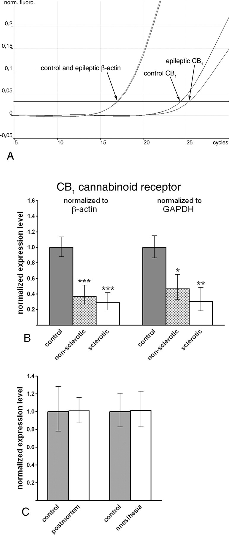
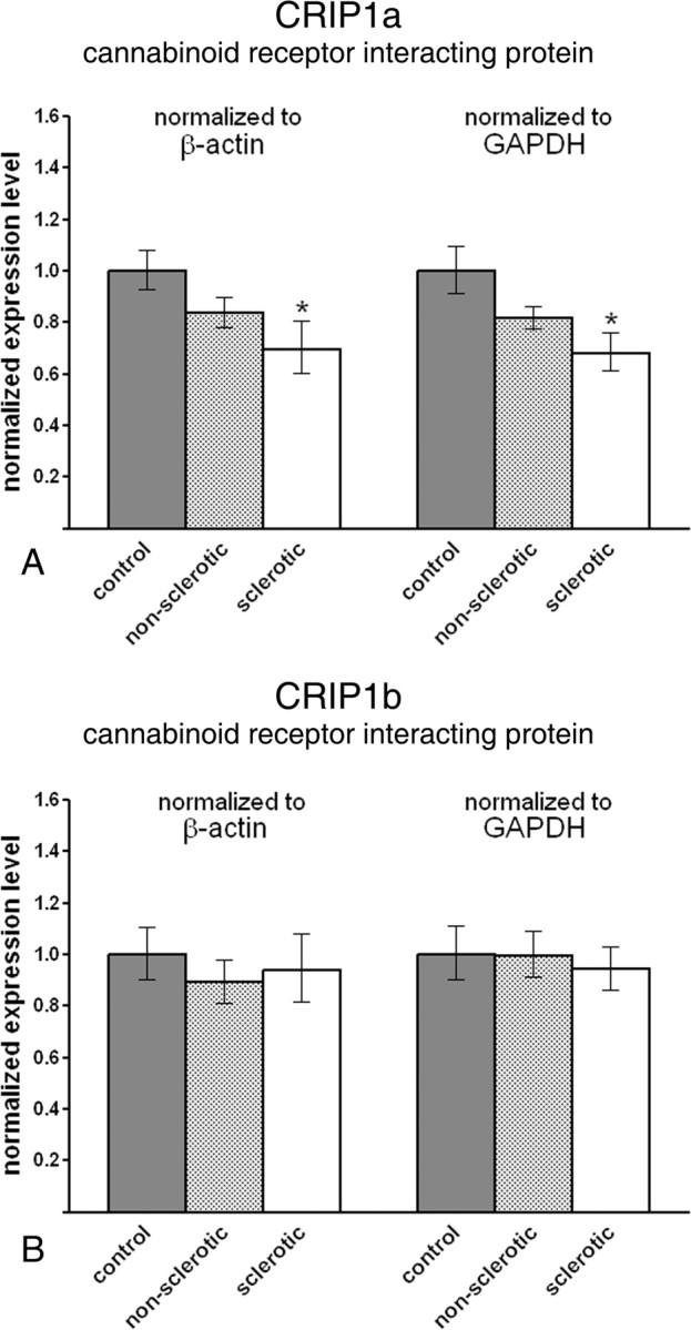
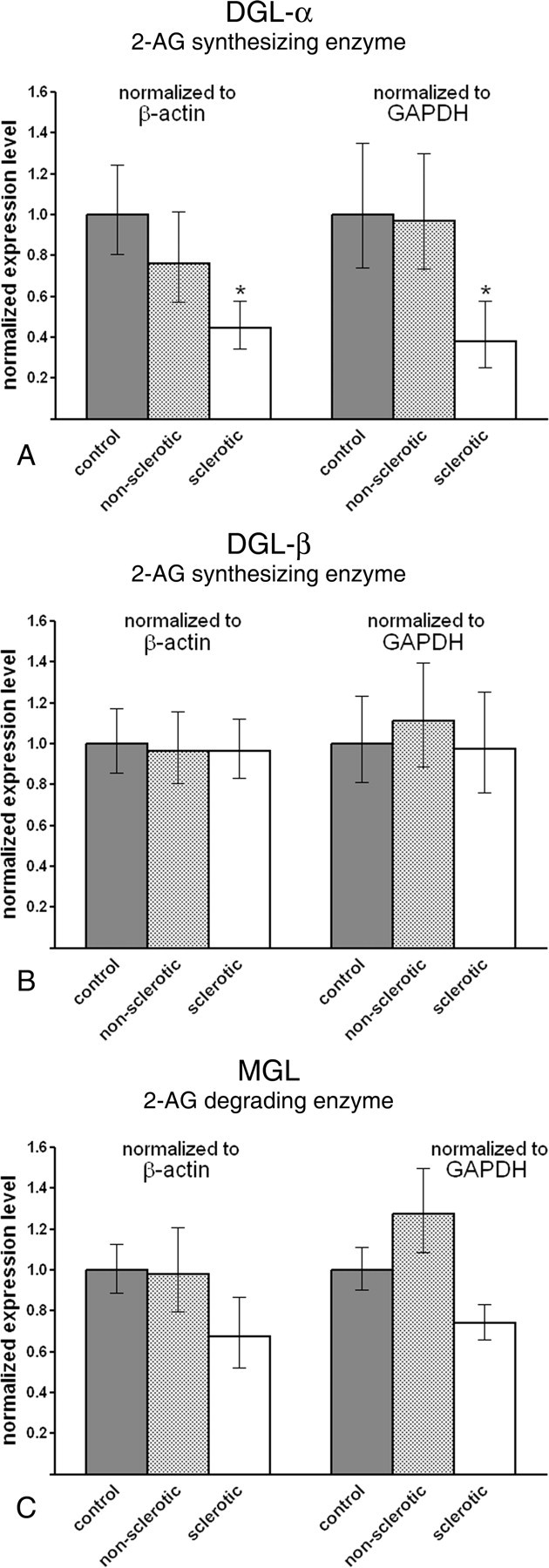
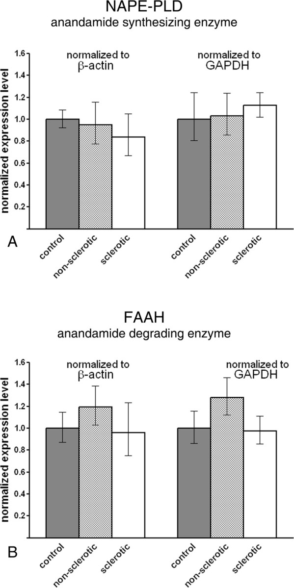
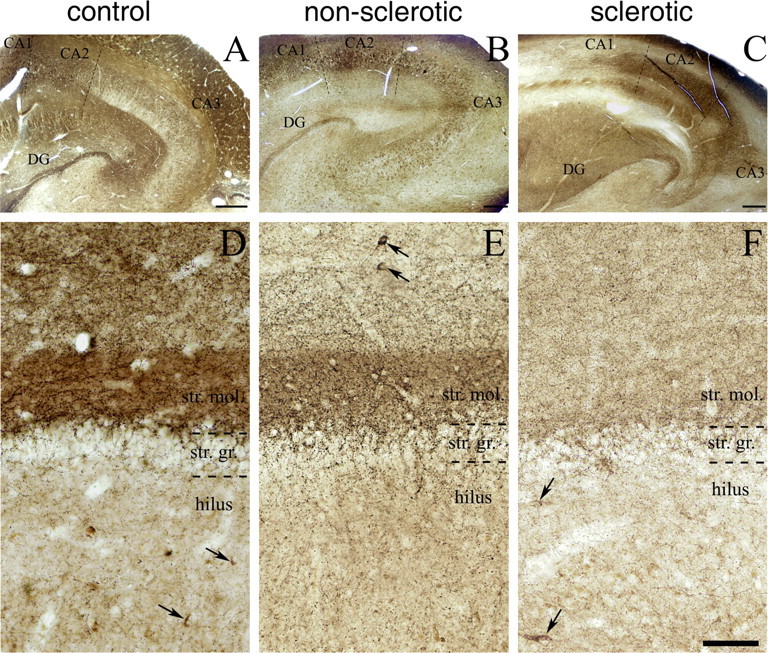
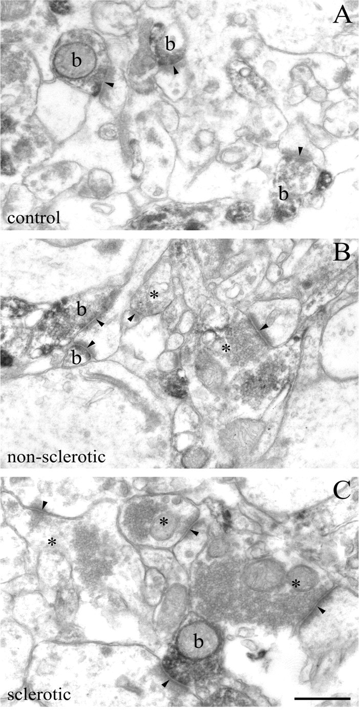
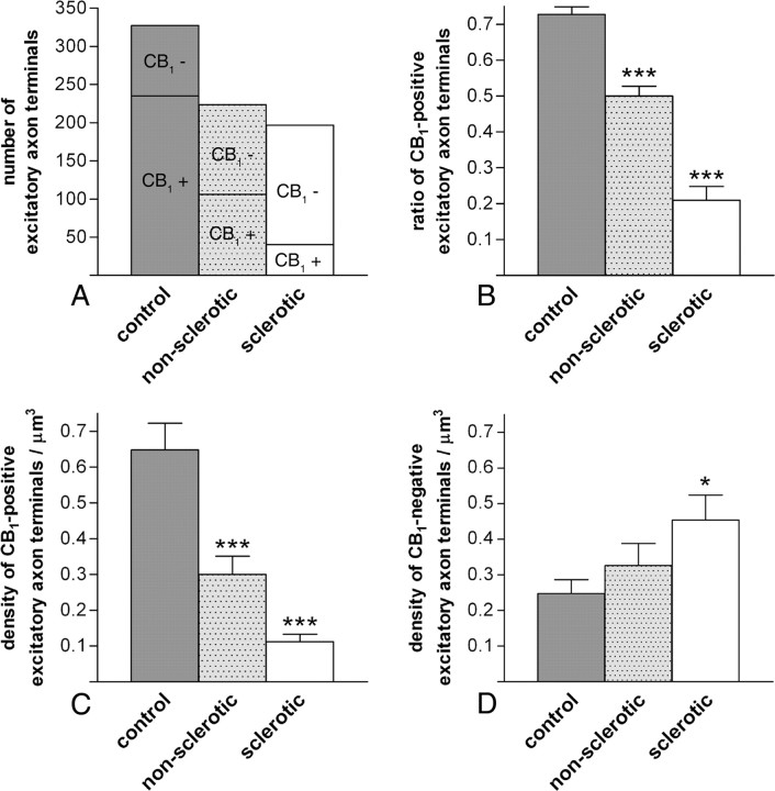
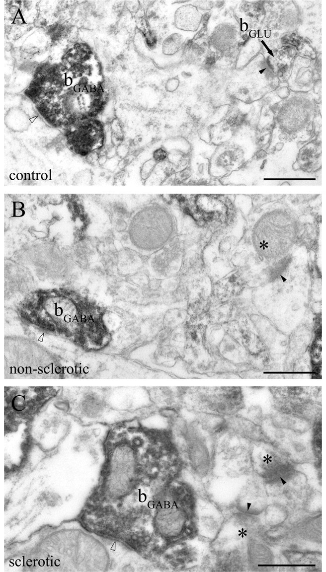
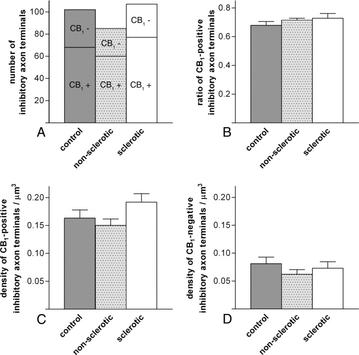
Endocannabinoid signaling is a key regulator of synaptic neurotransmission throughout the brain. Compelling evidence shows that its perturbation leads to development of epileptic seizures, thus indicating that endocannabinoids play an intrinsic protective role in suppressing pathologic neuronal excitability. To elucidate whether long-term reorganization of endocannabinoid signaling occurs in epileptic patients, we performed comparative expression profiling along with quantitative electron microscopic analysis in control (postmortem samples from subjects with no signs of neurological disorders) and epileptic (surgically removed from patients with intractable temporal lobe epilepsy) hippocampal tissue. Quantitative PCR measurements revealed that CB(1) cannabinoid receptor mRNA was downregulated to one-third of its control value in epileptic hippocampus. Likewise, the cannabinoid receptor-interacting protein-1a mRNA was decreased, whereas 1b isoform levels were unaltered. Expression of diacylglycerol lipase-alpha, an enzyme responsible for 2-arachidonoylglycerol synthesis, was also reduced by approximately 60%, whereas its related beta isoform levels were unchanged. Expression level of N-acyl-phosphatidylethanolamine-hydrolyzing phospholipase D and fatty acid amide hydrolase, metabolic enzymes of anandamide, and 2-arachidonoylglycerol's degrading enzyme monoacylglycerol lipase did not change. The density of CB(1) immunolabeling was also decreased in epileptic hippocampus, predominantly in the dentate gyrus, where quantitative electron microscopic analysis did not reveal changes in the ratio of CB(1)-positive GABAergic boutons, but uncovered robust reduction in the fraction of CB(1)-positive glutamatergic axon terminals. These findings show that a neuroprotective machinery involving endocannabinoids is impaired in epileptic human hippocampus and imply that downregulation of CB(1) receptors and related molecular components of the endocannabinoid system may facilitate the deleterious effects of increased network excitability.


Figures

Figure 1.
Figure 1.
CB1 receptor mRNA level is downregulated in the epileptic human hippocampus. A, Representative real-time PCR measurement of CB1 cannabinoid receptor mRNA level in control and epileptic human hippocampus. Note that the housekeeping gene β-actin reaches threshold of normalized fluorescence intensity at identical values in both the control and epileptic hippocampus. In contrast, when CB1 cannabinoid receptor mRNA is measured, the exponential phase begins later and reaches threshold approximately one cycle later in a representative sample from the epileptic hippocampus. One cycle difference in the cycle threshold value indicates ∼50% difference in the original mRNA level because of the exponential nature of the PCR. B, Gene expression level of the CB1 receptor is robustly downregulated in both nonsclerotic (n = 7) and sclerotic (n = 6) epileptic hippocampi compared with control tissue (n = 7). Note that the direction and magnitude of expression level changes were identical in parallel experiments using two distinct housekeeping genes, β-actin and GAPDH. C, Bar graphs demonstrate that neither postmortem delay nor sustained anesthesia influence CB1 mRNA level in the mouse hippocampus. Data are presented as mean expression ratio ± SEM. *p < 0.05; **p < 0.01; ***p < 0.001.
Figure 2.
Figure 2.
Gene expression level of cannabinoid receptor-interacting protein CRIP1a, but not CRIP1b, is reduced in the sclerotic epileptic hippocampus. A, CRIP1a mRNA level is significantly decreased in the sclerotic epileptic hippocampus (n = 6) compared with control values (n = 7). In contrast, the decrease in the nonsclerotic hippocampus (n = 7) did not reach significance. The direction and magnitude of expression level changes in the sclerotic hippocampus were identical in parallel experiments using two distinct housekeeping genes, β-actin and GAPDH. B, In contrast, CRIP1b mRNA levels did not differ significantly between nonsclerotic and sclerotic epileptic hippocampi compared with control values. Data are presented as mean expression ratio ± SEM. *p < 0.05.
Figure 3.
Figure 3.
Gene expression level of DGL-α, the biosynthetic enzyme of 2-AG, is diminished in the sclerotic epileptic hippocampus. A, DGL-α mRNA level is decreased to one-half of its control level in the sclerotic epileptic hippocampus. In contrast, significant difference in mRNA level was not observed between the nonsclerotic hippocampal samples and control subjects. Importantly, the direction and magnitude of expression level changes were identical in parallel experiments using two distinct housekeeping genes, β-actin and GAPDH. B, The related isoenzyme DGL-β is unaffected in the epileptic hippocampus. Real-time PCR measurement did not reveal significant changes in mRNA level either in the nonsclerotic or in the sclerotic epileptic hippocampus as compared with control values. C, MGL, the enzyme responsible for elimination of 2-AG, showed a slight but insignificant decrease in mRNA level in the sclerotic hippocampus [normalized to β-actin (p = 0.171) or to GAPDH (p = 0.079)]. In the nonsclerotic hippocampal samples, there was no indication of any subtle changes in gene expression level. Data are presented as mean expression ratio ± SEM. *p < 0.05.
Figure 4.
Figure 4.
Metabolic enzymes of anandamide are not downregulated in the epileptic hippocampus. A, Real-time PCR measurement did not reveal alterations in the gene expression level of NAPE-PLD, a key synthetic enzyme of anandamide. The normalized expression level was similar in all three experimental groups and in the parallel experiments, which used β-actin or GAPDH as reference genes. B, FAAH, the degrading enzyme of anandamide, did not show significant expression changes in the epileptic hippocampal samples. Data are presented as mean expression ratio ± SEM.
Figure 5.
Figure 5.
Immunostaining for CB1 cannabinoid receptor is reduced in the hippocampus of epileptic patients, particularly in the inner molecular layer of the dentate gyrus (DG). A, Light micrograph illustrating profound CB1 immunoreactivity throughout the human hippocampal formation of control subjects. By using a highly sensitive guinea pig antibody for CB1, the immunostaining highlights the different layers and subfields of the hippocampus according to the spatial arrangements of excitatory pathways. B, C, Although the general pattern of CB1 immunostaining is similar in the nonsclerotic and sclerotic epileptic hippocampi, the density of CB1 immunoreactivity is reduced in several layers. D–F, The most striking differences between the control and the epileptic hippocampi are visible in the dentate gyrus. The very dense neuropil-like labeling in the inner third of stratum moleculare (str. mol.) is evident in the control sample (D), but it is less so in the nonsclerotic epileptic sample (E), and it disappears almost completely in the sclerotic epileptic samples (F). In contrast, the stratum granulosum (str. gr.) and the hilus remained similar in all three experimental groups. Scattered cell bodies of GABAergic interneurons were also stained for CB1 (labeled by arrows), but conversely, there was no striking difference either in their distribution pattern or in their number between the control and epileptic human samples. Scale bars: A–C, 500 μm; D–F (in F), 100 μm.
Figure 6.
Figure 6.
Density of glutamatergic axon terminals bearing presynaptic CB1 cannabinoid receptors is decreased in the epileptic human hippocampus. A–C, The electron micrograph demonstrates a robust accumulation of strong CB1 immunoreactivity within axon terminals in the inner third of the stratum moleculare of control subjects. These CB1-positive boutons (b) form the classic asymmetric synapses (arrowheads) with an extensive postsynaptic density on dendritic spine heads. In control samples, nearly all axon terminals with asymmetric synapses are positive for CB1, whereas in the nonsclerotic (B) and sclerotic (C) samples, the number of CB1-positive asymmetric synapses drops noticeably. Note that a lack of staining does not necessarily mean the complete absence of CB1 receptors, but it means that the antigen level fails to reach detection threshold in these CB1-negative boutons (depicted by asterisks). Scale bar, 0.5 μm.
Figure 7.
Figure 7.
Quantitative analysis of the ratio and density of CB1-positive excitatory axon terminals in the inner molecular layer of the human dentate gyrus. A, The number of excitatory axon terminals either positive or negative for CB1 cannabinoid receptor was established using an unbiased stereological estimation method (Geinisman et al., 1996). Altogether, 1092 disector pairs were analyzed, which resulted in 327 terminals in control, 224 terminals in nonsclerotic epileptic, and 197 terminals in sclerotic epileptic patients. The number of CB1-positive terminals decreased from 235 terminals in control subjects to 106 or 40 terminals in the nonsclerotic or sclerotic epileptic samples, respectively. In contrast, the number of CB1-negative terminals increased from 92 terminals in control subjects to 118 or 157 terminals in nonsclerotic or sclerotic epileptic samples, respectively. In the quantitative analysis, tissue samples from three individuals from each experimental group were used. B, The ratio of CB1-positive excitatory axon terminals versus all excitatory axon terminals was 72.8 ± 2.1% in control, 50 ± 2.8% in nonsclerotic epileptic, and 21 ± 3.8% in sclerotic epileptic samples (mean ± SEM). The difference between control and epileptic samples was highly significant (χ2 test, ***p < 0.001 both for nonsclerotic and sclerotic epileptic samples). C, The estimated numerical density of CB1-positive axon terminals in the inner molecular layer of the dentate gyrus of control subjects (0.648 ± 0.075/μm3) was strongly decreased in nonsclerotic epileptic patients (0.3 ± 0.051/μm3) and in sclerotic epileptic patients (0.112 ± 0.021/μm3) as well (values are mean ± SEM). This sharp decline in the density of CB1-positive axon terminals was statistically significant (ANOVA, p < 0.001). Significance exists between control values and both nonsclerotic and sclerotic epileptic patients (Dunnett's post hoc test, ***p < 0.001). D, The estimated numerical density of CB1-negative axon terminals was elevated in epileptic samples [density in control samples, 0.248 ± 0.038/μm3; in nonsclerotic samples, 0.326 ± 0.062/μm3; and in sclerotic samples, 0.454 ± 0.07/μm3 (mean ± SEM)]. Increase was significant between analyzed groups (ANOVA, p = 0.04); density of CB1-negative axon terminals in sclerotic epileptic patients was significantly increased compared with control values (Dunnett's post hoc test, *p = 0.037), but not in nonsclerotic patients (Dunnett's post hoc test, p = 0.546).
Figure 8.
Figure 8.
CB1-positive GABAergic axon terminals are intact in the epileptic human hippocampus. A–C, The electron micrographs show striking CB1 immunoreactivity within GABAergic axon terminals (bGABA) forming symmetric synapses (open arrowheads) in the inner third of stratum moleculare of the dentate gyrus. Dense accumulation of the end product of immunoperoxidase reaction (DAB) indicates that these GABAergic axon terminals are fully equipped with CB1 receptors in both the control and the epileptic hippocampi. Glutamatergic boutons terminate on dendritic spine heads with typical asymmetric synapses (closed arrowheads), which is characterized by broad postsynaptic density. In the control sample, the glutamatergic axon terminal (bGLU) is positive for CB1 (A), whereas CB1-negative boutons (asterisks) forming asymmetric synapses are shown in electron micrographs taken from the nonsclerotic (B) and sclerotic (C) samples. Note that CB1-positive axon terminals forming inhibitory synapses are larger than those giving excitatory synapses. Scale bars, 0.5 μm.
Figure 9.
Figure 9.
Quantitative analysis of the ratio and density of CB1-positive inhibitory axon terminals in the inner molecular layer of the human dentate gyrus. A–D, The ratio and density of inhibitory axon terminals either positive or negative for CB1 cannabinoid receptor were established in a manner similar to that detailed in Figure 7 for excitatory terminals. A, Altogether, 1092 disector pairs were analyzed, which resulted in 102 inhibitory terminals in control, 85 terminals in nonsclerotic epileptic, and 107 terminals in sclerotic epileptic patients. The number of CB1-positive terminals forming symmetric synapses was 68 terminals in control subjects, and 60 or 77 terminals in the nonsclerotic or sclerotic epileptic samples, respectively. The number of CB1-negative terminals was 34 terminals in control subjects, and 25 or 30 terminals in nonsclerotic or sclerotic epileptic samples, respectively. B, The ratio of CB1-positive GABAergic axon terminals versus all GABAergic axon terminals was 67.8 ± 2.7% in control, 71.5 ± 1.3% in nonsclerotic epileptic, and 72.9 ± 3.3% in sclerotic epileptic samples (mean ± SEM). The difference between control and epileptic samples was not significant (χ2 test, p = 0.698). C, The estimated numerical density of CB1-positive inhibitory axon terminals in the inner molecular layer of the dentate gyrus of control subjects (0.16 ± 0.02/μm3) was similar in nonsclerotic epileptic patients (0.15 ± 0.01/μm3) and in sclerotic epileptic patients (0.19 ± 0.02/μm3) (ANOVA, p = 0.114). D, The estimated numerical density of CB1-negative inhibitory axon terminals was comparable in the three groups (density in control samples, 0.08 ± 0.01/μm3; in nonsclerotic samples, 0.06 ± 0.01/μm3; and in sclerotic samples, 0.07 ± 0.01/μm3; ANOVA, p = 0.458).

Similar articles

Cited by

References

    1. Alger BE. Retrograde signaling in the regulation of synaptic transmission: focus on endocannabinoids. Prog Neurobiol. 2002;68:247–286. - PubMed
    1. Amaral DG. A Golgi study of cell types in the hilar region of the hippocampus in the rat. J Comp Neurol. 1978;182:851–914. - PubMed
    1. Ameri A, Wilhelm A, Simmet T. Effects of the endogeneous cannabinoid, anandamide, on neuronal activity in rat hippocampal slices. Br J Pharmacol. 1999;126:1831–1839. - PMC - PubMed
    1. Becker AJ, Chen J, Paus S, Normann S, Beck H, Elger CE, Wiestler OD, Blumcke I. Transcriptional profiling in human epilepsy: expression array and single cell real-time qRT-PCR analysis reveal distinct cellular gene regulation. NeuroReport. 2002;13:1327–1333. - PubMed
    1. Bernard C, Milh M, Morozov YM, Ben-Ari Y, Freund TF, Gozlan H. Altering cannabinoid signaling during development disrupts neuronal activity. Proc Natl Acad Sci USA. 2005;102:9388–9393. - PMC - PubMed

Publication types

MeSH terms

LinkOut - more resources