. 2008 Mar 19;28(12):3060-70.
doi: 10.1523/JNEUROSCI.5450-07.2008.

Synaptic metaplasticity through NMDA receptor lateral diffusion

Affiliations

Synaptic metaplasticity through NMDA receptor lateral diffusion

Jiang Zhao et al. J Neurosci. .

Retraction in

Abstract

Lateral diffusion of glutamate receptors was proposed as a mechanism for regulating receptor numbers at synapses and affecting synaptic functions, especially the efficiency of synaptic transmission. However, a direct link between receptor lateral diffusion and change in synaptic function has not yet been established. In the present study, we demonstrated NMDA receptor (NMDAR) lateral diffusion in CA1 neurons in hippocampal slices by detecting considerable recovery of spontaneous or evoked EPSCs from the block of (+)-MK-801 [(+)-5-methyl-10,11-dihydro-5H-dibenzo [a,d] cyclohepten-5,10-imine maleate], an irreversible NMDAR open-channel blocker. We observed changes on both the number and the composition of synaptic NMDAR on recovery. More importantly, after the recovery, long-term potentiation (LTP)-producing protocol induced only LTD (long-term depression) instead of LTP. In contrast, a complete recovery from competitive NMDAR blocker D,L-AP-5 was observed without subsequent changes on synaptic plasticity. Our data suggest a revised model of NMDAR trafficking wherein extrasynaptic NMDARs, mostly NR1/NR2B receptors, move laterally into synaptic sites, resulting in altered rule of synaptic modification. Thus, CA1 synapses exhibit a novel form of metaplasticity in which the direction of synaptic modification can be reverted through subtype-specific lateral diffusion of NMDA receptors.


Figures

Figure 1.
Figure 1.
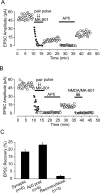
Anomalous recovery of NMDAR-mediated evoked EPSCs from MK-801 block. A, Representative traces show progressive block of AMPAR- and NMDAR-mediated components in an averaged evoked EPSC trace administrated with NBQX (5 μm) and MK-801 (10 μm), respectively. All the recordings were conducted in whole-cell configuration in low-Mg2+ (0.25 mm) ACSF with the GABAA antagonist BMI (10 μm). B, Paired-pulse stimuli (100 ms interpulse interval) evoked NMDAR-mediated EPSCs. The recording was conducted in low-Mg2+ ACSF with both GABAA and AMPA receptor antagonists. In our recording, the second evoked EPSCs were always bigger than the first ones. The paired-pulse ratio was measured as the ratio of the second current amplitude over the first current amplitude. C, Selective block of synaptic NMDAR-mediated EPSCs. Because paired-pulse stimuli could facilitate presynaptic release of glutamate, which in turn facilitates the activation of synaptic NMDAR currents, 50 paired-pulse stimuli were delivered at 0.125 Hz with simultaneous MK-801 perfusion, which could selectively block opened synaptic NMDAR channels. However, during 30 min washout period, we observed a progressive gradual EPSC recovery and finally recovered up to ∼35% of the control. D, Agonist-evoked block of NMDAR-mediated EPSCs. Whole-cell application of NMDA (1 mm) through puffer in the presence of the noncompetitive NMDA receptor open channel blocker MK-801 (10 μm) almost completely abolish evoked EPSCs. After a 30 min washout period, no significant recovery was observed. Circle in the inset refers to the boundary between synaptic and extrasynaptic region. The cylinder refers to the effective blocking area by MK-801.
Figure 2.
Figure 2.
The recovery of EPSCs from irreversible MK-801 block suggests adding of new receptors through lateral diffusion. A, The competitive NMDA blocker AP-5 did not prevent recovery of evoked EPSCs, suggesting that the recovery was not attributable to MK-801 unbinding. After MK-801 was washed out, the averaged EPSC amplitude was measured before and after 10 min AP-5 (100 μm) block. After removal of AP-5, the extent of recovery was even higher than that before AP-5 application. B, Irreversible block of NMDAR-mediated EPSCs by whole-cell coapplication of NMDA and MK-801 after EPSC recovery from MK-801 block suggested that unbinding of receptors with lower affinity for MK-801 was not responsible for the recovery. D, AP-5 was added in the first recovery period to match the protocol used in A, as was previously conducted by Tovar and Westbrook (2002). C, Statistical graph shows partial recovery of EPSCs after synaptic stimulation (Synaptic), postblock application of AP-5 (Ap5 post; measured as the difference of recovery level before and after AP-5 application in A), and coapplication of NMDA and MK-801 after recovery from AP-5 block (Recovery/block).
Figure 3.
Figure 3.
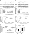
Increased recovery level of NMDAR-mediated spontaneous EPSCs in perforated patch mode suggests the possible involvement of intracellular components in recovery. A, Selective block of synaptic NMDAR-mediated spontaneous EPSCs recorded in conventional whole-cell patch mode. Top, Representative traces from neurons at the indicated experiment conditions (control, paired-pulse stimulation/MK801, recovery). Bottom, Spontaneous NMDAR EPSCs recorded at different time points before and after selective synaptic NMDARs block through paired-pulse stimuli given at 0.125 Hz accompanied by MK-801 application. After 30 min MK-801 washout, partial recovery of EPSCs was observed. B, Agonist-evoked block of NMDAR-mediated spontaneous EPSCs recorded in conventional whole-cell patch mode. Top, Representative traces from neurons at the indicated experiment conditions (control, NMDA/MK801, recovery). Bottom, Complete and irreversible block of spontaneous NMDAR current after whole-cell coapplication of agonist NMDA and MK-801. C, Cumulative distribution of sEPSC amplitude (mean ± SEM) before MK-801 application (control) and after recovery from MK-801 (recovery) recorded in conventional whole-cell patch mode (left) and perforated patch mode (right), respectively. D, Partial recovery of NMDAR-mediated sEPSC frequency in two recording modes. Normalized sEPSC frequency over different time points show significantly increased recovery level in perforated patch mode than that in conventional patch mode. E, Statistical graphs show partial recovery of sEPSC amplitude and frequency in two recording modes, with frequency recovered up to 36.65 ± 5.63% of the initial level in conventional patch mode compared with 67.55 ± 6.22% in perforated patch mode (*p < 0.05). Amplitude recovery was 65.12 ± 6.46% in conventional patch mode compared with 73.85 ± 7.13% in perforated patch mode (no significance).
Figure 4.
Figure 4.
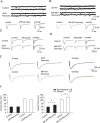
NR2 subunit composition switches after NMDAR-mediated EPSC recovery from MK-801 block. A, No change on deactivation constant (decay time) of spontaneous NMDAR-mediated EPSCs after recovery from AP-5 block. Unless stated in text, most of following data were obtained in perforated patch configuration. Top, Representative spontaneous recordings before and after recovery from AP-5 block. Bottom, Averaged spontaneous traces show no change in decay time. B, Prolongation of decay time of spontaneous NMDAR-mediated EPSCs after recovery from MK-801 block. Data are from neurons with full frequency recovery. Top, Representative spontaneous recordings before and after recovery from MK-801 block. Bottom, Averaged spontaneous traces display increase of decay time. C, Representative traces display no significant change in sensitivity of spontaneous NMDAR-mediated EPSCs to the NR2B-selective antagonist ifenprodil (3 μm) before (top) and after (bottom) recovery from AP-5 (100 μm) block confirmed by overlay of two current curves (right row). D, Representative traces before (top) and after (bottom) recovery from MK-801 (10 μm) block display marked increase in sensitivity to ifenprodil confirmed by overlay of two current curves (right row). E, Prolongation of decay time after recovery from MK-801 was also observed on evoked NMDAR-mediated EPSCs recorded in low-magnesium ACSF (bottom). In contrast, no alteration was observed on decay time of evoked EPSCs after AP-5 recovery (top). F, Statistical graphs show significant elevation on decay time of spontaneous and evoked EPSCs after recovery from MK-801 (right) and show no change on decay time of EPSCs after AP-5 recovery (left) (*p < 0.05).
Figure 5.
Figure 5.
Differential regulation of synaptic plasticity after recovery from MK-801 and AP-5 block. A, After full recovery of sEPSCs from AP-5 block, standard LTP-producing stimulus (2 Hz, 200 pulses during a 2.5 min depolarization to 0 mV) can induce LTP of AMPAR-mediated EPSCs. Left of the vertical dashed line, NMDAR-mediated sEPSCs at different time points before and after recovery from AP-5. Full recovery of both sEPSC amplitude and frequency were observed. Right of the dashed line, Evoked EPSC (0.125 Hz) before and after a brief LTP-producing stimulation. B, Conversion of synaptic plasticity direction from LTP to LTD after partial recovery from MK-801 block. After partial recovery of EPSC amplitude and full recovery of EPSC frequency from MK-801 (10 μm) block, the same LTP-producing stimulus protocol (2 Hz, 200 pulses during a 2.5 min depolarization to 0 mV) induced LTD instead of LTP of AMPAR-mediated EPSCs. To confirm that the LTD is not attributable to deterioration of the cell recorded, AP-5 was applied at the end of recording and LTD was abolished, which demonstrated that the observed LTD was dependent on NMDAR activity. C and D are statistical plots showing the whole recording process delineated in A and B, respectively. n = 7 for C and D. Representative traces from indicated times are presented above the graphs. E, In the control condition with only paired-pulse stimulation, neither change of sEPSC nor conversion from LTP to LTD was observed. Representative traces from indicated times are presented above the graphs.
Figure 6.
Figure 6.
Conversion of synaptic plasticity direction is not attributable to partial inhibition of NMDAR function after recovery from MK-801 block. A, The graph displays progressive recovery of evoked NMDAR-mediated EPSCs from MK-801 block recorded in perforated patch mode. The representative averaged traces from indicated times are presented above the graph. B, AP-5 at a concentration of 5–8 μm, which could reduce NMDAR-mediated EPSCs to a level similar to that observed after recovery from MK-801 block, did not induce conversion of LTP to LTD but abolished LTP induction. The representative averaged traces of evoked EPSCs from different time points of control and AP-5 block are presented above the graph.
Figure 7.
Figure 7.
Partial NR2B inhibition blocks conversion of synaptic plasticity direction after recovery from MK-801 block. A, Partial NR2B inhibition by a low concentration of ifenprodil (0.6 μm) blocks conversion of synaptic plasticity direction. Top, Sample of evoked NMDAR-mediated currents showing partial inhibition by the NR2B antagonist ifenprodil (0.6 μm), which elevates the NR2A/NR2B ratio to close to control level. Bottom, After partial recovery of sEPSC amplitude and full recovery of sEPSC frequency from MK-801 (10 μm) block, a low concentration of ifenprodil (0.6 μm) was immediately applied, and an STP instead of LTD was induced with LTP-producing stimulus protocol (2 Hz, 200 pulses during a 2.5 min depolarization to 0 mV). B, Similar NMDAR EPSC amplitude inhibition by AP-5 at a concentration of 8–10 μm could not block conversion of synaptic plasticity direction. Top, Sample of evoked NMDAR-mediated currents showing similar partial inhibition by AP-5 (8–10 μm) as ifenprodil (0.6 μm) did. Bottom, After partial recovery of sEPSC amplitude and full recovery of sEPSC frequency from MK-801 (10 μm) block, a low concentration of AP-5 (8–10 μm) was immediately applied, and LTD was still induced with LTP-producing stimulus protocol (2 Hz, 200 pulses during a 2.5 min depolarization to 0 mV).
Figure 8.
Figure 8.
Field potential recordings confirm differential regulation of synaptic plasticity after recovery from MK-801 and AP-5 block. A, Consecutive LTP induced by two tetanic stimuli (100 Hz, twice, 120 s interval). LTP was produced by first tetanic stimulus and the second LTP was further induced on the base of the first one by the second tetanic stimulus. The interval of the two titanic stimuli was 40 min. B, Recovery of LTP induction after removal of AP-5. LTP induction was blocked by AP-5 perfusion, but, after 30 min AP-5 washout, LTP induction could be completely recovered with tetanic stimulation. The extent of field EPSP potentiation was similar to that produced by the first tetanic stimulus in control (A). C, Conversion of synaptic plasticity direction from LTP to LTD after partial recovery from MK-801 block. After 30 min MK-801 washout, the same LTP-producing stimulus protocol induced LTD instead of LTP, indicating conversion of synaptic plasticity direction. D, Superimposed data show differential regulation of synaptic plasticity after recovery from MK-801 and AP-5 block.

Similar articles

Cited by

References

    1. Abraham WC, Bear MF. Metaplasticity: the plasticity of synaptic plasticity. Trends Neurosci. 1996;19:126–130. - PubMed
    1. Adesnik H, Nicoll RA, England PM. Photoinactivation of native AMPA receptors reveals their real-time trafficking. Neuron. 2005;48:977–985. - PubMed
    1. Bartlett TE, Bannister NJ, Collett VJ, Dargan SL, Massey PV, Bortolotto ZA, Fitzjohn SM, Bashir ZI, Collingridge GL, Lodge D. Differential roles of NR2A and NR2B-containing NMDA receptors in LTP and LTD in the CA1 region of two-week old rat hippocampus. Neuropharmacology. 2007;52:60–70. - PubMed
    1. Bear MF. Bidirectional synaptic plasticity: from theory to reality. Philos Trans R Soc Lond B Biol Sci. 2003;358:649–655. - PMC - PubMed
    1. Bekkers JM, Stevens CF. Presynaptic mechanism for long-term potentiation in the hippocampus. Nature. 1990;346:724–729. - PubMed

Publication types

MeSH terms