. 2008 May 15;111(10):5152-62.
doi: 10.1182/blood-2007-10-116889. Epub 2008 Mar 19.

BH3-only proteins Noxa, Bmf, and Bim are necessary for arsenic trioxide-induced cell death in myeloma

Affiliations

BH3-only proteins Noxa, Bmf, and Bim are necessary for arsenic trioxide-induced cell death in myeloma

Alejo A Morales et al. Blood. .

Abstract

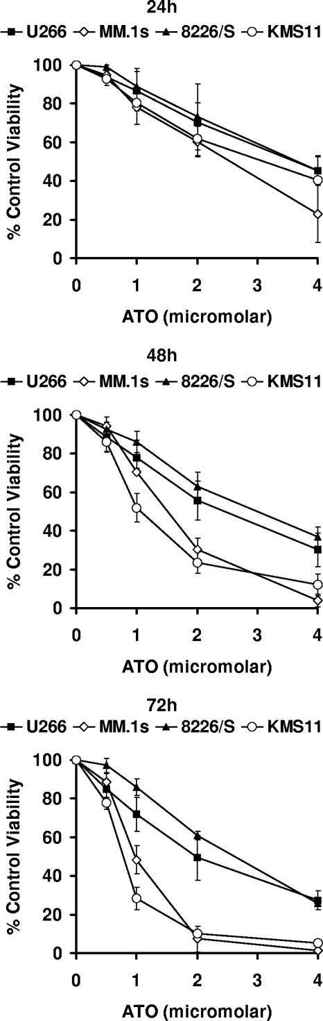
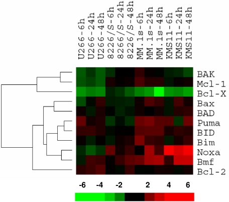
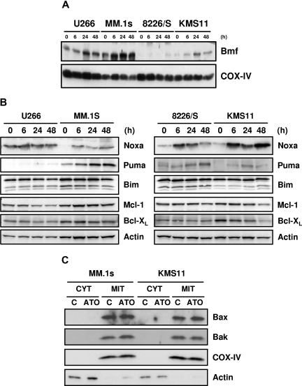
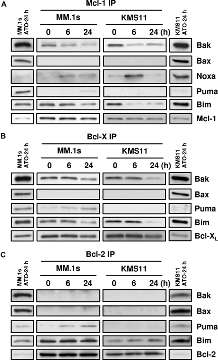
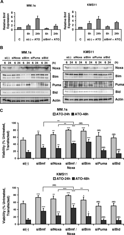
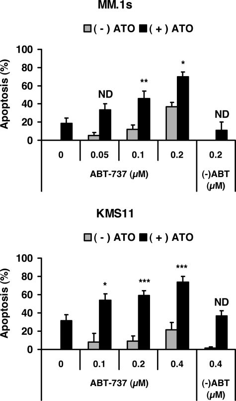
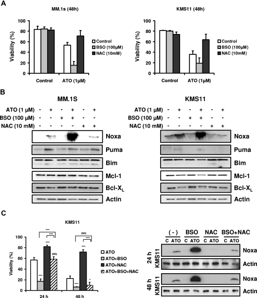
The use of arsenic trioxide (ATO) to treat multiple myeloma (MM) is supported by preclinical studies as well as several phase 2 studies, but the precise mechanism(s) of action of ATO has not been completely elucidated. We used gene expression profiling to determine the regulation of apoptosis-related genes by ATO in 4 MM cell lines and then focused on Bcl-2 family genes. ATO induced up-regulation of 3 proapoptotic BH3-only proteins (Noxa, Bmf, and Puma) and down-regulation of 2 antiapoptotic proteins Mcl-1 and Bcl-X(L). Coimmunoprecipitation demonstrated that Noxa and Puma bind Mcl-1 to release Bak and Bim within 6 hours of ATO addition. Bak and Bim are also released from Bcl-X(L). Silencing of Bmf, Noxa, and Bim significantly protected cells from ATO-induced apoptosis, while Puma silencing had no effect. Consistent with a role for Noxa inhibition of Mcl-1, the Bad-mimetic ABT-737 synergized with ATO in the killing of 2 MM lines. Finally, Noxa expression was enhanced by GSH depletion and inhibited by increasing GSH levels in the cells. Understanding the pattern of BH3-only protein response should aid in the rational design of arsenic-containing regimens.


Figures

Figure 1
Figure 1
ATO-induced apoptosis in 4 human myeloma cell lines. U266, MM.1s, 8226/S, and KMS11 were treated for 24, 48, and 72 hours with 0, 0.5, 1, 2, and 4 μM ATO. Cell viability was determined by flow cytometry following annexin-V–FITC/PI staining. Percentage (%) of control viability was plotted versus ATO concentration. Graphs are presented for each time point. The data are presented as the means (± SD) of 3 independent experiments.
Figure 2
Figure 2
Gene expression profile for ATO-induced changes in the Bcl-2 family. U266, MM.1s, 8226/S, and KMS11 cell lines were treated with 2 μM ATO for 6, 24, and 48 hours. Total RNA was obtained and cRNA was probed on Affymetrix Hu133 2.0 Plus Chips. Signal intensity was used as gene expression. Ratios at 6, 24, and 48 hours versus baseline gene expression were calculated for each probe set and average ratios were obtained for genes. Bcl-2 family member genes were clustered using the bioinformatic programs Cluster and TreeView. The bar at the bottom represents the scale for fold changes.
Figure 3
Figure 3
Up-regulation of Bmf, Noxa, and Puma and down-regulation of Mcl-1 and Bcl-XL by ATO. U266, MM.1s, 8226/S, and KMS11 were treated for 6, 24, and 48 hours with 2 μM ATO. (A) Mitochondrial-rich fractions were obtained and Bmf expression was determined by Western blot. Membranes were reprobed with rabbit anti–COX IV polyclonal antibody to determine loading. (B) Western blot analysis of total protein lysates from ATO-treated cells. Membranes were probed with specific antibodies for Noxa, Puma, Bim, Mcl-1, Bcl-XL, and actin. (C) Cytosolic and heavy membrane fractions were obtained for untreated and 24-hour ATO-treated MM.1s and KMS11 cells as indicated in “Subcellular fractionation.” Actin and COX IV were used as indicators of fraction purity.
Figure 4
Figure 4
Coimmunoprecipitation of proapoptotic proteins with Mcl-1, Bcl-XL, and Bcl-2. MM.1s and KMS11 cell lines were treated with 2 μM ATO for 0, 6, and 24 hours. Protein lysates were prepared using 2% CHAPS buffer. (A) Mcl-1, (B) Bcl-XL, and (C) Bcl-2 proteins were immunoprecipitated with mAbs. Coimmunoprecipitated proteins were detected by Western blot using specific antibodies for Bak, Bax, Noxa, Bim, Puma, Mcl-1, Bcl-XL, and Bcl-2. The separated bands on each end of the panels represent the input for the IP. This lane contains 10% of the input, however only 60% of the immunoprecipitated protein was loaded in the gel. All bands represented are from the same experiment and same exposure of film.
Figure 5
Figure 5
BH3-only proteins Bmf, Noxa, and Bim silencing protected cells from ATO-induced apoptosis. MM.1s and KMS11 cell lines were electroporated with siBmf, siNoxa, siBim, siPuma, and siBid (SmartPool; DHARMACON RNA Technologies). Nontargeting siRNA (si(-)) pool was used as a negative control. After 16 hours, cells were treated with 2 μM ATO for 6 and 24 hours for real-time PCR or Western blot analysis and for 24 and 48 hours for ATO-induced apoptosis analysis. (A) Total RNA was obtained for si(-) control and siBmf samples treated with ATO. Real-time PCR was used to determine Bmf transcript expression. Bmf relative expression refers to Bmf transcript expression relative to GAPDH transcript expression. (B) Protein lysates were obtained for si(-) control–, siNoxa-, siBim-, siPuma-, and siBid-transfected cells and silencing was determined by Western blot at 6 and 24 hours after ATO treatment. (C) siRNA electroporated cells were treated with 2 μM ATO for 24 and 48 hours. Viability was evaluated by annexin-V–FITC/PI staining. Percentage (%) of control (untreated, transfected) viability was plotted versus time (hours). The data are presented as the means (± SD) of 4 and 5 independent experiments for MM.1s and KMS11, respectively. t test was used to compare differences among samples, si(-), and experimental combinations unless otherwise indicated, with confidence intervals of 95%. ND indicates no difference; *P < .05; **P < .01; ***P < .001.
Figure 6
Figure 6
Effect of Bcl-2/Bcl-XL inhibitor on ATO-induced apoptosis. MM.1s and KMS11 cell lines were treated with 2 μM ATO and indicated concentrations of ABT-737 or the less active enantiomer (-)ABT for 24 hours. Viability was measured by annexin-V–FITC/PI staining. Percentage (%) of apoptosis was plotted versus the concentration of ABT-737. The data are presented as the means (± SD) of 5 independent experiments. t test was used to compare differences between expected additive effect and actual cotreatment results, with confidence intervals of 95%. ND indicates no difference; *P < .05; **P < .01; ***P < .001.
Figure 7
Figure 7
BSO sensitized cells to ATO and enhanced up-regulation of the BH3-only protein Noxa. (A) MM.1s and KMS11 cells were treated with 1 μM ATO as a single agent or in combination with BSO (100 μM) or NAC (10 mM) for 48 hours. Viability was evaluated by annexin-V–FITC/PI staining. Data are presented as means (± SD) of 3 independent experiments. (B) MM.1s and KMS11 cells were treated for 24 hours. Protein expression was determined by Western blot. Membranes were reprobed with specific antibodies for Noxa, Puma, Bim, Mcl-1, Bcl-XL, and actin. (C) KMS11 cells were treated with 2 μM ATO as a single agent or in combination with BSO and/or NAC for 24 and 48 hours. Viability was evaluated by annexin-V–FITC/PI staining. Data are presented as means (± SD) of 4 independent experiments. Noxa expression was determined by Western blot. t test was used to compare differences among samples, ATO, and experimental combinations unless otherwise indicated, with confidence intervals of 95%. ND indicates no difference; *P < .05; **P < .01; ***P < .001.

Similar articles

Cited by

References

    1. Jemal A, Siegel R, Ward E, et al. Cancer statistics, 2006. CA Cancer J Clin. 2006;56:106–130. - PubMed
    1. Hideshima T, Leif Bergsagel P, Kuehl WM, Anderson KC. Advances in biology of multiple myeloma: clinical applications. Blood. 2004;104:607–618. - PubMed
    1. Rousselot P, Labaume S, Marolleau JP, et al. Arsenic trioxide and melarsoprol induce apoptosis in plasma cell lines and in plasma cells from myeloma patients. Cancer Res. 1999;59:1041–1048. - PubMed
    1. Park WH, Seol JG, Kim ES, et al. Arsenic trioxide-mediated growth inhibition in MC/CAR myeloma cells via cell cycle arrest in association with induction of cyclin-dependent kinase inhibitor, p21, and apoptosis. Cancer Res. 2000;60:3065–3071. - PubMed
    1. Grad JM, Bahlis NJ, Reis I, Oshiro MM, Dalton WS, Boise LH. Ascorbic acid enhances arsenic trioxide-induced cytotoxicity in multiple myeloma cells. Blood. 2001;98:805–813. - PubMed

Publication types

MeSH terms