

Coronary flow velocity reserve and carotid intima media thickness in patients with autosomal dominant polycystic kidney disease: from impaired tubules to impaired carotid and coronary arteries
- PMID: 18354076
- PMCID: PMC2440264
- DOI: 10.2215/CJN.02330607
Coronary flow velocity reserve and carotid intima media thickness in patients with autosomal dominant polycystic kidney disease: from impaired tubules to impaired carotid and coronary arteries
Abstract
Background and objectives: Cardiovascular problems are a major cause of morbidity and mortality in patients with autosomal dominant polycystic kidney disease. Endothelial dysfunction, an early and reversible feature in the pathogenesis of atherosclerosis, is associated with increased vascular smooth muscle tone, arterial stiffening, and increased intima-media thickness. Coronary flow velocity reserve is a noninvasive test showing endothelial function of epicardial coronary arteries and coronary microcirculatory function. The aim of the study was to investigate the carotid intima-media thickness and coronary flow velocity reserve in patients with autosomal dominant polycystic kidney disease.
Design, setting, participants, & measurements: Thirty normotensive patients with autosomal dominant polycystic kidney disease (10 male, 20 female) with well-preserved renal function and 30 healthy subjects (12 male, 18 female) were included in the study. Coronary flow velocity reserve was measured at baseline and after dipyridamole infusion by echocardiography. Coronary flow velocity reserve was calculated as the ratio of hyperemic to baseline diastolic peak velocities.
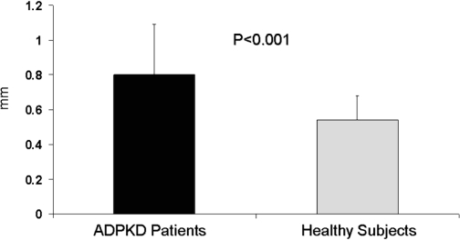
Results: Carotid intima-media thickness was significantly higher in patients than in control subjects (0.80 +/- 0.29 versus 0.54 +/- 0.14 mm, respectively; P < 0.001). Moreover, coronary flow velocity reserve was significantly lower in patients than in control subjects (1.84 +/- 0.39 versus 2.65 +/- 0.68, respectively; P < 0.001).
Conclusions: Normotensive patients with autosomal dominant polycystic kidney disease with well-preserved renal function have significantly increased carotid intima-media thickness and significantly decreased coronary flow velocity reserve compared with healthy subjects. These findings suggest that atherosclerosis starts at an early stage in the course of their disease in patients with autosomal dominant polycystic kidney disease.
Figures
References
-
- Gabow PA: Autosomal dominant polycystic kidney disease. N Engl J Med 329: 332–342, 1993 - PubMed
-
- Iglesias CG, Torres VE, Offord K, Holley KE, Beard CM, Kurland LT: Epidemiology of adult polycystic kidney disease, Olmsted County, Minnesota: 1935–1980. Am J Kidney Dis 2: 630–639, 1983 - PubMed
-
- Fick GM, Johnson AM, Hammond WS, Gabow PA: Causes of death in autosomal dominant polycystic kidney disease. J Am Soc Nephrol 5: 2048–2056, 1995 - PubMed
-
- Ecder T, Schrier RW: Hypertension in autosomal dominant polycystic kidney disease: Early occurrence and unique aspects. J Am Soc Nephrol 12: 194–200, 2001 - PubMed
-
- Schrier RW, McFann KK, Johnson AM: Epidemiological study of kidney survival in autosomal dominant polycystic kidney disease. Kidney Int 63: 678–685, 2003 - PubMed
Publication types
MeSH terms
LinkOut - more resources
Full Text Sources
Medical
