. 2008 Apr 9;27(7):1029-38.
doi: 10.1038/emboj.2008.53. Epub 2008 Mar 20.

The fission yeast homologue of CENP-B, Abp1, regulates directionality of mating-type switching

Affiliations

The fission yeast homologue of CENP-B, Abp1, regulates directionality of mating-type switching

Lorena Aguilar-Arnal et al. EMBO J. .

Abstract

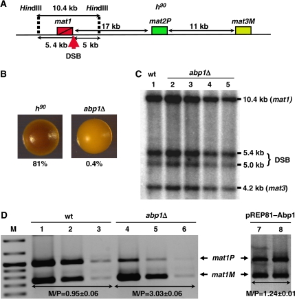
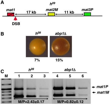
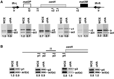
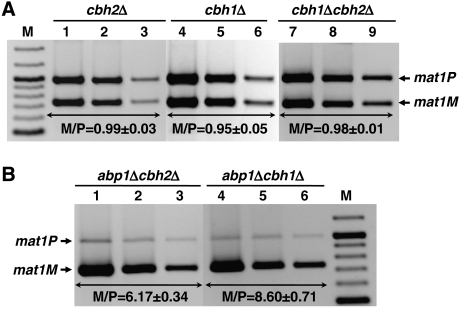
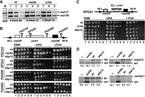
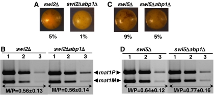
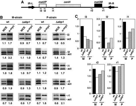
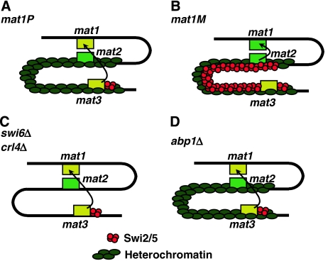
In fission yeast, mating-type switching involves replacing genetic information contained at the expressed mat1 locus by that of either the mat2P or mat3M donor loci. Donor selection is nonrandom, as mat1P cells preferentially use mat3M for switching, whereas mat1M cells use mat2P. Switching directionality is determined by the cell-type-specific distribution of the Swi2-Swi5 complex that, in mat1P cells, localises to mat3M and, only in mat1M cells, spreads to mat2P in a heterochromatin-dependent manner. Mechanisms regulating spreading of Swi2-Swi5 across heterochromatin are not fully understood. Here, we show that the fission yeast homologue of CENP-B, Abp1, binds to the silent domain of the mating-type locus and regulates directionality of switching. Deletion of abp1 prevents utilisation of mat2P, as when heterochromatin is disrupted and spreading of Swi2-Swi5 is impaired. Our results show that, indeed, deletion of abp1 abolishes spreading of Swi2-Swi5 to mat2P. However, in abp1Delta cells, heterochromatin organisation at the mating-type locus is preserved, indicating that Abp1 is actually required for efficient spreading of Swi2-Swi5 through heterochromatin. Cbh1 and Cbh2, which are also homologous to CENP-B, have only a minor contribution to the regulation of directionality of switching, which is in contrast with the strong effects observed for Abp1.


Figures

Figure 1
Figure 1
Analysis of the effects on mating-type switching of deleting abp1 in a homothallic h90 strain. (A) Schematic representation of the structural organisation of the mating-type region in an h90 strain. The positions of mat1, mat2P and mat3M loci are indicated. The position of the SSB/DSB imprint, contained within the 10.4-kb HindIII fragment spanning the mat1 locus, is also indicated. (B) Iodine staining of wild-type (h90) and abp1Δ colonies. Before staining, colonies were grown in sporulation medium at 25°C for 3 days. Numbers below each panel indicate the frequencies of sporulation. Similar results were obtained with several independent abp1Δ colonies. (C) Presence of the SSB/DSB imprint was determined by Southern blot analysis of a wild-type (lane 1) and four independent abp1Δ strains (lanes 2–5). Genomic DNA was digested with HindIII and probed with a 9-kb DNA fragment, which spans the mat1 locus and contains mat3M information at mat1, so that it also detects a 4.2-kb HindIII band corresponding to the mat3M locus. Bands arising from cleavage at the SSB/DBS imprint of the 10.4-kb HindIII fragment are indicated (DSB). (D) Quantitative multiplex PCR determination of the predominant mating type adopted by wild-type (wt, lanes 1–3) and abp1Δ cells (lanes 4–6). PCR reactions were performed using appropriate primers to amplify mat1M and mat1P sequences simultaneously. For each strain, three 10-fold dilutions of the PCR products were analysed. Similar results were obtained with several independent abp1Δ colonies. Lanes 7 and 8, correspond to abp1Δ cells transformed with plasmid pREP81–Abp1 to express Abp1. In this case, single dilutions of the PCR products obtained from two independent isolates are shown. The positions of the bands corresponding to mat1P and mat1M are indicated. The M/P ratios (±s.d.) are indicated below the corresponding lanes.
Figure 2
Figure 2
Analysis of the effects on mating-type switching of deleting abp1 in h09 cells. (A) Schematic representation of the structural organisation of the mating-type region in an h09 strain. The positions of mat1, mat2M and mat3P loci are indicated. The position of the SSB/DSB imprint is also indicated. (B) Iodine staining of wild-type (h09) and abp1Δ colonies. Numbers below each panel correspond to the frequencies of sporulation. (C) Quantitative multiplex PCR determination of the predominant mating type adopted by wild-type h09 (lanes 1–3) and abp1Δ cells (lanes 4–6). PCR reactions were performed as in Figure 1D. The M/P ratios (±s.d.) are indicated below the corresponding lanes.
Figure 3
Figure 3
Abp1 localises to the silent domain of the mating-type locus. (A) Binding of Abp1–HA at the silent domain of the mating-type locus was determined by ChIP-analysis using αHA antibodies. Immunoprecipitated material was analysed by multiplex PCR using primers specific for the indicated regions of the mating-type locus (bands ir, r2, c1, c2, l1 and l2) (see Supplementary Table SII for a description of the primers used) and, in the same PCR reaction, primers that amplify a fragment of similar length of the act1 locus (bands act), used as control. (B) Similar experiments as those described in (A) but with primers designed to amplify shorter DNA fragments flanking region l1 (bands p1, p2 and p4) or contained within it (bands p3). In this case, a shorter fragment from the act1 gene was used as control (bands act(s)). Lanes WCE correspond to PCR products obtained from the input material before immunoprecipitation. Lanes αHA correspond to the products obtained from the immunoprecipitated material. Numbers below each lane correspond to the ratio of the corresponding mating-type-specific band with respect to the control act1 band.
Figure 4
Figure 4
Analysis of the effects on mating-type switching of deleting cbh1 and cbh2 in h90 cells. (A) Quantitative multiplex PCR determination of the predominant mating type adopted by cbh2Δ (lanes 1–3), cbh1Δ (lanes 4–6) and cbh1Δcbh2Δ cells (lanes 7–9). (B) Quantitative multiplex PCR determination of the predominant mating type adopted by abp1Δcbh2Δ (lanes 1–3) and abp1Δcbh1Δ cells (lanes 4–6). PCR reactions were performed as in Figure 1D. The M/P ratios (±s.d.) are indicated below the corresponding lanes.
Figure 5
Figure 5
Heterochromatin organisation of the mating-type region is not disrupted in abp1Δ cells. (A) Quantitative multiplex PCR determination of the predominant mating type adopted by wild-type (lanes 1–3), swi6Δ (lanes 4–6) and crl4Δ cells (lanes 7–9). PCR reactions were performed as in Figure 1D. The M/P ratios (±s.d.) are indicated below the corresponding lanes. (B) The effect of deleting abp1 on heterochromatin-mediated silencing was determined in several strains carrying ura4+ insertions at different positions of the silent domain of the mating-type locus. Sites of insertion are schematically indicated on top. Exponentially growing cells were plated as serial 10-fold dilutions (lanes 1–6) onto selective media with (panels EMM) or without uracil (panels −URA), or in the presence of FOA (panels +FOA). For each strain, several independent abp1Δ isolates were analysed and they all showed very similar results. (C) Similar results as in (B) but performed with strain SPG27, where the entire cenH element was replaced by ura4+. In this case, the results obtained with four independent abp1Δ isolates are presented. (D) Swi6 deposition at the silent domain of the mating-type region was determined in wild-type (lanes wt) and abp1Δ cells (lanes abp1Δ) by ChIP analysis using αSwi6 antibodies. Immunoprecipitated material was analysed as in Figure 3 with primers to amplify specific bands of the IR-R and IR-L regions (bands IRr and IR), the cenH element (bands cenH/c1) and a region just left of mat3 (bands mat3/r2) (see Supplementary Table SII for a description of the primers used). Lanes swi6Δ correspond to immunoprecipitation experiments performed with αSwi6 antibodies in a swi6Δ strain, missing Swi6. Lanes – correspond to mock immunoprecipitation experiments in which no antibodies were added. Numbers below each lane correspond to the ratio of the corresponding mating-type-specific band with respect to the control act band.
Figure 6
Figure 6
Analysis of the effects on mating-type switching of deleting abp1 in swi2Δ (A, B) and swi5Δ (C, D) cells. (A, C) Iodine staining of the indicated strains. Numbers below each panel correspond to the frequencies of sporulation. (B, D) Quantitative multiplex PCR determination of the predominant mating type adopted by each of the indicated strains. PCR reactions were performed as in Figure 1D. The M/P ratios (±s.d.) are indicated below the corresponding lanes.
Figure 7
Figure 7
Deletion of abp1 impairs spreading of Swi2–Swi5 to mat2. The effects of deleting abp1 on the distribution of Swi2–myc along the silent domain of the mating-type locus were determined in both a stable M-strain (mat1smto) and a stable P-strain (mat1PΔ17) by ChIP analysis using α-myc antibodies. (A) Regions of the silent domain of the mating-type locus where the presence of Swi2–myc was analysed (see Supplementary Table SII for a description of the primers used). (B) ChIP analysis corresponding to wild-type (wt) and abp1Δ cells derived from either a stable M-strain (panel M-strain) or a stable P-strain (panel P-strain). Material obtained after immunoprecipitation with α-myc antibodies was analysed as described in Figure 3 using primers specific for the indicated regions (bands r1, r2, l1, l2 and l3). Lanes WCE correspond to PCR products obtained from the input material before immunoprecipitation. Lanes α-myc correspond to the products obtained from the immunoprecipitated material. Numbers below each lane correspond to the ratio of the corresponding mating-type-specific band with respect to the control act band. (C) Quantitative analysis of the results shown in (B). For each of the mating-type-specific regions analysed, the relative fold of enrichment of the corresponding band in the immunoprecipitated material with respect to the input material (WCE) is presented for both wild type (wt) and abp1Δ cells in a stable M or P background.
Figure 8
Figure 8
Model to account for the contribution of Abp1 to the regulation of directionality of mating-type switching. In mat1P cells, the Swi2–Swi5 complex localises to mat3 (A) and, only in mat1M cells, spreads to mat2 (B), allowing its use as a donor during switching. Spreading of Swi2–Swi5 to mat2 is mediated by heterochromatin being abolished by mutations in heterochromatin assembly factors (i.e. swi6Δ and crl4Δ), which prevent use of mat2 as a donor (C). In abp1Δ cells, heterochromatin organisation of the mating-type locus is preserved but spreading of Swi2–Swi5 to mat2 is impaired and, as a consequence, mat2 is not efficiently used as a donor during switching (D).

References

    1. Akamatsu Y, Dziadkowiec D, Ikeguchi M, Shinagawa H, Iwasaki H (2003) Two different Swi5-containing protein complexes are involved in mating-type switching and recombination repair in fission yeast. Proc Natl Acad Sci USA 100: 15770–15775 - PMC - PubMed
    1. Arcangioli B (1998) A site- and strand-specific DNA break confers asymmetric switching potential in fission yeast. EMBO J 10: 4503–4510 - PMC - PubMed
    1. Arcangioli B, Klar AJS (1991) A novel switch-activating site (SAS1) and its cognate binding factor (SAP1) required for efficient mat1 switching in Schizosaccharomyces pombe. EMBO J 10: 3025–3032 - PMC - PubMed
    1. Arcangioli B, Thon G (2004) Mating-type cassettes: structure, switching and silencing. In The Molecular Biology of Schizosaccharomyces pombe, Egel R (ed), pp 129–144. Berlin, Heidelberg, New York: Springer-Verlag
    1. Bähler J, Wu J-Q, Longtine MS, Shah NG, McKenzie A III, Steever AB, Wach A, Philippsen P, Pringle JR (1998) Heterologous modules for efficient and versatile PCR-based gene targeting in Schizosaccharomyces pombe. Yeast 14: 943–951 - PubMed

Publication types

MeSH terms