. 2008 Apr 23;27(8):1266-76.
doi: 10.1038/emboj.2008.52. Epub 2008 Mar 20.

The initiation factor eIF3-f is a major target for atrogin1/MAFbx function in skeletal muscle atrophy

Affiliations

The initiation factor eIF3-f is a major target for atrogin1/MAFbx function in skeletal muscle atrophy

Julie Lagirand-Cantaloube et al. EMBO J. .

Abstract

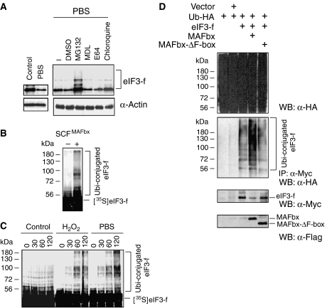
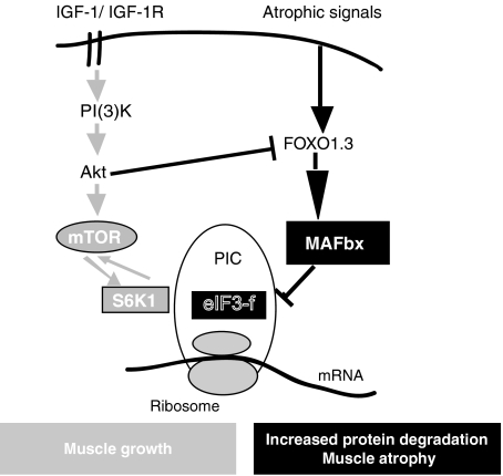
In response to cancer, AIDS, sepsis and other systemic diseases inducing muscle atrophy, the E3 ubiquitin ligase Atrogin1/MAFbx (MAFbx) is dramatically upregulated and this response is necessary for rapid atrophy. However, the precise function of MAFbx in muscle wasting has been questioned. Here, we present evidence that during muscle atrophy MAFbx targets the eukaryotic initiation factor 3 subunit 5 (eIF3-f) for ubiquitination and degradation by the proteasome. Ectopic expression of MAFbx in myotubes induces atrophy and degradation of eIF3-f. Conversely, blockade of MAFbx expression by small hairpin RNA interference prevents eIF3-f degradation in myotubes undergoing atrophy. Furthermore, genetic activation of eIF3-f is sufficient to cause hypertrophy and to block atrophy in myotubes, whereas genetic blockade of eIF3-f expression induces atrophy in myotubes. Finally, eIF3-f induces increasing expression of muscle structural proteins and hypertrophy in both myotubes and mouse skeletal muscle. We conclude that eIF3-f is a key target that accounts for MAFbx function during muscle atrophy and has a major role in skeletal muscle hypertrophy. Thus, eIF3-f seems to be an attractive therapeutic target.


Figures

Figure 1
Figure 1
Interaction of the E3 ubiquitin ligase MAFbx with eIF3-f. (A) Interaction of MAFbx and eIF3-f. GST or GST–MAFbx beads (2 μg) were incubated with extracts (200 μg) of C2C12 myoblasts overexpressing Myc-tagged eIF3-f. After washing resins, the bound proteins were eluted, subjected to SDS–PAGE along with an extract sample and analysed by immunoblotting with anti-Myc antibody. (B) Co-immunoprecipitation of exogenously expressed MAFbx and eIF3-f. C2C12 myotubes were cotransfected with Myc-tagged eIF3-f and Flag-tagged MAFbx expression constructs. Total cellular extracts (250 μg) were subjected to immunoprecipitation with anti-Myc and/or anti-Flag, followed by immunoblotting analysis with anti-Flag and/or anti-Myc antibodies. (C) eIF3-f colocalizes in the nucleus in the presence of MAFbx. C2C12 myotubes were cotransfected with mammalian expression plasmids encoding GFP–MAFbx and Myc–eIF3-f and, after 24 h, the transfected cells were fixed and immunostained with anti-Myc antibodies and DAPI. Images of a representative field were obtained by indirect immunofluorescence microscopy. Scale bar, 50 μm. (D) Confocal fluorescence imaging of endogenous eIF3-f during starvation-induced atrophy in C2C12 myotubes. Myotubes were fixed 6 h after food deprivation and stained for DNA (blue), eIF3-f (green, upper panels) and MAFbx (green, lower panels). Scale bar, 50 μm. Arrows indicate nuclear localization of eIF3-f and MAFbx during starvation-induced atrophy.
Figure 2
Figure 2
Glucocorticoid treatment, starvation of cells and oxidative stress induced upregulation of MAFbx and degradation of eIF3-f. (A) Myotubes at 3 days of differentiation were treated with 225 μM H2O2 and 1, 10 and 50 μM DEX for 24 h or were starved by removal of growth medium, amino acids and glucose and incubated in PBS. Proteins were extracted and subjected to immunoblot analysis with specific antibodies against MAFbx and eIF3-f. α-Tubulin is shown as a loading control. (B) Expression of eIF3-f mRNA was analysed by semiquantitative RT–PCR. Myotubes were treated with increasing concentrations of DEX for 24 h (1, 10 and 50 μM), or incubated either in PBS for 6 h and medium was replaced in refed cultures for 12 h or in H2O2 (225 μM) for 4 and 9 h. GAPDH expression was used as an internal control. (C) Depletion of MAFbx affects eIF3-f degradation in C2C12 myotubes undergoing atrophy. C2C12 myoblasts were transfected with pTER+MAFbx or pTER+control. Myotubes at 2 days of differentiation were treated with H2O2 (225 μM) or were incubated in PBS and myotubes were harvested 6 h later. Cell lysates were prepared and total proteins were subjected to western blot with anti-eIF3-f and anti-MAFbx antibodies. (D) Opposite expression of MAFbx and eIF3-f in the skeletal muscle of mice during starvation and refeeding. Shown is an immunoblot analysis of protein extracts from hind limb muscles, using anti-eIF3-f, anti-MAFbx and anti-tropomyosin antibodies. Mice were starved for 48 h (starved) and refed for 48 h (Refed). Control mice were fed randomly.
Figure 3
Figure 3
Food deprivation- and oxidative stress-induced atrophy increases polyubiquitination and degradation of eIF3-f by the SCFMAFbx pathway. (A) C2C12 myotubes at day 4 of differentiation were incubated in PBS for 4 h in the presence of DMSO, MG132 (30 μM), MDL (50 μM), E64 (30 μM) and chloroquine (40 μM). Fifty micrograms of total proteins was subjected to western blotting with anti-eIF3-f antibodies. Anti-α-tubulin antibodies were used as an internal control. (B) The recombinant SCFMAFbx complex was assayed for the ability to mediate polyubiquitination of eIF3-f in the presence of E1, ATP, ubiquitin and cdc34 (E2). Reaction mixtures were separated on SDS–PAGE, followed by autoradiography. (C) Myotubes at 4 days of differentiation were treated with 225 μM H2O2 (9 h) and/or were starved by removal of growth medium and incubated in PBS (6 h). Cell lysates were prepared and 30 μg of total proteins was used for in vitro ubiquitination assay. 35S-labelled eIF3-f was subjected to in vitro ubiquitination reaction in the presence of normal and/or atrophic C2C12 myotube lysates. Reaction mixtures were separated on SDS–PAGE, followed by autoradiography. (D) MAFbx increases eIF3-f ubiquitination in vivo. C2C12 myotubes were transiently transfected with expression plasmids encoding Myc-eIF3-f, HA–ubiquitin, Flag–MAFbx and/or the F-box mutant Flag–MAFbx-ΔF-box. Transfected cells were treated for 2 h with 50 μM MG132 before harvesting. Cells were lysed in denaturation buffer containing 0.5% SDS. Aliquots (10%) were analysed by western blotting with anti-HA antibodies (upper panel). Cell lysates were then diluted in immunoprecipitation buffer, subjected to immunoprecipitation with anti-Myc antibodies (middle panel) and analysed by western blotting with anti-Flag, anti-Myc and anti-HA antibodies (lower panels).
Figure 4
Figure 4
MAFbx increases eIF3-f turnover. C2C12 myoblasts were cotransfected using the expression vector encoding Myc–eIF3-f and Flag-tagged MAFbx-wt or the mutant MAFbx-ΔF-box as indicated. The cells were treated with CHX 36 h after transfection to block protein synthesis. Cell lysates were prepared at the indicated time and analysed by immunoblotting. Quantification of eIF3-f turnover following CHX treatment based on densitometric scanning of two experiments is shown. MG132 was added to control eIF3-f degradation by the proteasome.
Figure 5
Figure 5
Genetic repression of eIF3-f in differentiated myotubes induces atrophy but genetic activation of eIF3-f is sufficient to block starvation-induced atrophy. (A) Scheme of the protocol applied to C2C12 cells to obtain transfected populations of myotubes. Myoblasts were transfected using Lipofectamin 2000 and cultured for 3 days in GM and then in DM for 6 days. During the first 5 days of culture, Tetra was added and then it was omitted until starvation-induced atrophy. (B) eIF3-f expression accounts for the atrophic and hypertrophic response of myotubes. Transfected cells were green fluorescent by virtue of GFP in the pBI bicistronic expression vector (images in the right column) in a field of myotubes (phase-contrast images in the left column). Scale bar, 50 μm. (C) Transiently transfected C2C12 myoblasts were induced to differentiate in the presence of Tetra for 3 days and then in the absence of Tetra for 3 more days. Myotubes were starved by removal of growth medium, amino acids and glucose and incubated in PBS for 7 h. Total cellular proteins (50 μg) were analysed by immunoblotting. Expression of eIF3-f sense constructs was confirmed by western blotting with anti-HA antibodies. The endogenous eIF3-f expression levels were analysed by western blotting with the polyclonal anti-eIF3-f. Anti-Cdk4 antibodies were used as loading controls. The graph represents averaged densitometric quantification of the data from three replicate experiments. *P<0.03. (D) Myotube diameter after food starvation for 4 and 7 h in the absence of Tetra. Experiments were repeated twice with similar results. Data represent the average±s.e. of at least 120 myotubes. *P<0.05. (E) Myotubes were stained with Hoechst 33258 to visualize the nuclei. The average number of nuclei per myotubes is indicated. Data are means±s.e.m. of at least 100 myotubes. *P<0.05.
Figure 6
Figure 6
Overexpression of eIF3-f induces hypertrophy in muscle cells. (A) Clones of C2C12 myoblasts stably expressing conditional HA-tagged eIF3-f (MCK-eIF3-f) or empty vector (control) were differentiated for 3 days, fixed and stained with Hansen's hemalun. Images of a representative field were obtained by light microscopy. Scale bar, 100 μm. (B) Myotube diameter was measured at day 3 of differentiation. Histogram is a mean±s.e.m. of at least 125 myotubes. *P<0.05. (C) Clones of C2C12 myoblasts stably expressing conditional HA-tagged eIF3-f (MCK-eIF3-f) or empty vector (control) were cultured in GM (20% FCS) for 72 h and then in DM (2% FCS) for 96 h. Proteins were resolved on a 10% SDS–PAGE and HA–eIF3-f, endogenous eIF3-f and α-tubulin (as standard control) were detected by western blot analysis using specific antibodies. (D) Kinetics of differentiation of stably transfected C2C12 cells. Western blots with specific antibodies directed against myosin heavy chain, troponin T, desmin, MyoD and myogenin. * indicates a nonspecific band.
Figure 7
Figure 7
Overexpression of eIF3-f in muscle fibres induces hypertrophy. (A) Adult TA muscles were electroporated with HA-tagged eIF3-f (pMCK-HA-eIF3-f-IRES-GFP, right leg) or the empty vector (left leg) and mice were killed after 14 days. Hypertrophic fibres expressing eIF3-f were detected in transverse sections stained with anti-HA (eIF3-f) (right panel). Scale bar, 20 and 50 μm for merge pictures. (B) Histogram shows the fibre size distribution of TA from four mice. Black bars, vector control; grey bars, HA–eIF3-f. (C) Mean cross-sectional area of TA fibre from four mice ±s.e.m. **P<0.03 between control and the eIF3-f electrotransferred fibres, by Student's t-test (right panel). NS: nonsignificant.
Figure 8
Figure 8
Model depicting the two signalling pathways converging on the eIF3-f factor in the antagonism atrophy/hypertrophy of the skeletal muscle.

Similar articles

Cited by

References

    1. Aravind L, Ponting CP (1998) Homologues of 26S proteasome subunits are regulators of transcription and translation. Protein Sci 7: 1250–1254 - PMC - PubMed
    1. Asano K, Phan L, Anderson J, Hinnebusch AG (1998) Complex formation by all five homologues of mammalian translation initiation factor 3 subunits from yeast Saccharomyces cerevisiae. J Biol Chem 27: 18573–18585 - PubMed
    1. Baar K, Esser K (1999) Phosphorylation of p70(S6k) correlates with increased skeletal muscle mass following resistance exercise. Am J Physiol 276: C120–C127 - PubMed
    1. Bandyopadhyay A, Maitra U (1999) Cloning and characterization of the p42 subunit of mammalian translation initiation factor 3 (eIF3): demonstration that eIF3 interacts with eIF5 in mammalian cells. Nucleic Acids Res 27: 1331–1337 - PMC - PubMed
    1. Bodine SC, Latres E, Baumhueter S, Lai VK, Nunez L, Clarke BA, Poueymirou WT, Panaro FJ, Na E, Dharmarajan K, Pan ZQ, Valenzuela DM, DeChiara TM, Stitt TN, Yancopoulos GD, Glass DJ (2001a) Identification of ubiquitin ligases required for skeletal muscle atrophy. Science 294: 1704–1708 - PubMed

Publication types

MeSH terms

Substances