. 2008 Apr 9;27(7):1122-33.
doi: 10.1038/emboj.2008.50. Epub 2008 Mar 20.

Chloroplast monothiol glutaredoxins as scaffold proteins for the assembly and delivery of [2Fe-2S] clusters

Affiliations

Chloroplast monothiol glutaredoxins as scaffold proteins for the assembly and delivery of [2Fe-2S] clusters

Sibali Bandyopadhyay et al. EMBO J. .

Abstract

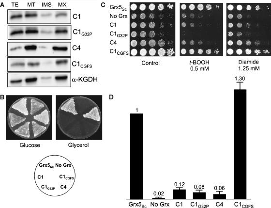
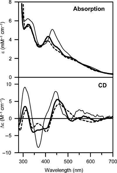
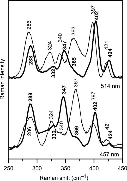
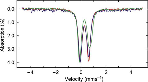
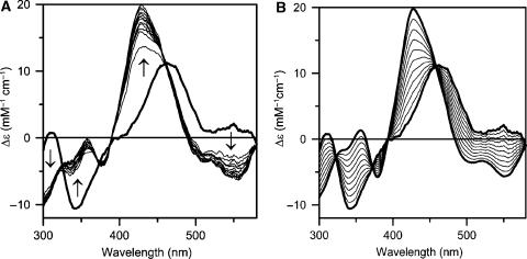
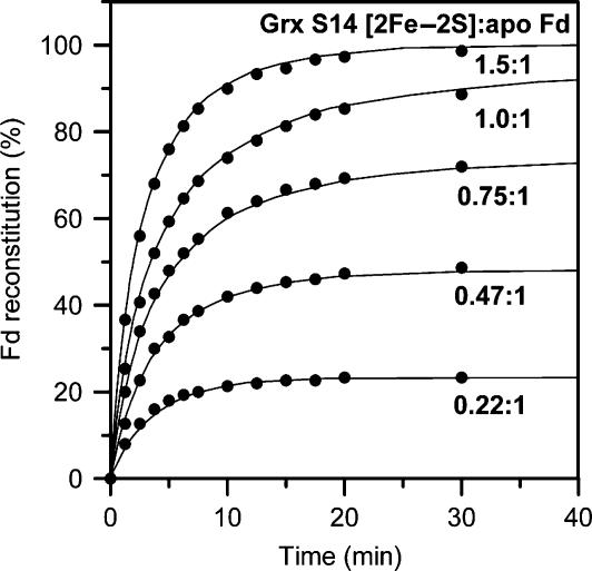
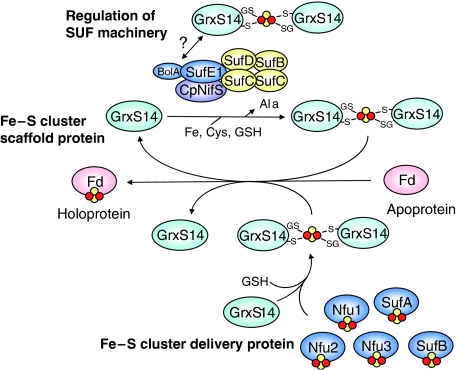
Glutaredoxins (Grxs) are small oxidoreductases that reduce disulphide bonds or protein-glutathione mixed disulphides. More than 30 distinct grx genes are expressed in higher plants, but little is currently known concerning their functional diversity. This study presents biochemical and spectroscopic evidence for incorporation of a [2Fe-2S] cluster in two heterologously expressed chloroplastic Grxs, GrxS14 and GrxS16, and in vitro cysteine desulphurase-mediated assembly of an identical [2Fe-2S] cluster in apo-GrxS14. These Grxs possess the same monothiol CGFS active site as yeast Grx5 and both were able to complement a yeast grx5 mutant defective in Fe-S cluster assembly. In vitro kinetic studies monitored by CD spectroscopy indicate that [2Fe-2S] clusters on GrxS14 are rapidly and quantitatively transferred to apo chloroplast ferredoxin. These data demonstrate that chloroplast CGFS Grxs have the potential to function as scaffold proteins for the assembly of [2Fe-2S] clusters that can be transferred intact to physiologically relevant acceptor proteins. Alternatively, they may function in the storage and/or delivery of preformed Fe-S clusters or in the regulation of the chloroplastic Fe-S cluster assembly machinery.


Figures

Figure 1
Figure 1
Subcellular localization of CGFS Grxs by GFP fusion. (A) GrxS14, (B) GrxS16 and (C) GrxS15. From left to right: visible light, autofluorescence of chlorophyll (red) or mitochondrial marker (white); fluorescence of the constructions and merged images. Only one of the guard cells shows chloroplast-localized GFP, because a small numbers of cells were transfected. As the mitochondrial marker (DsRed) is co-transfected with the GFP construction, it is only visible in the cell that expresses GFP.
Figure 2
Figure 2
Rescue of the S. cerevisiae grx5 mutant defects by poplar monothiol glutaredoxins. (A) Compartmentalization of GrxS14, S15, S16, S17 and S17398−492 in the mitochondrial matrix of S. cerevisiae cells. Cultures were grown exponentially in YPLactate medium at 30°C to about 3 × 10⁷ cells ml−1, before mitochondrial isolation and subfractionation. TE, total cell extract; MT, mitochondrial fraction; IMS, intermembrane space; MX, matrix. Proteins (20 μg) were loaded in the TE lanes, and 5 μg was loaded in the other lanes. Anti-HA anti-lipoic acid antibodies were used in the western blot to detect the HA-tagged proteins, and the matrix marker α-ketoglutarate dehydrogenase (α-KGDH). (B) Growth on glucose (YPD plates) or glycerol (YPGly plates), after 3 days at 30°C. (C) Sensitivity to t-BOOH or diamide of the strains after 3 days at 30°C on YPD plates. (D) Ratio between aconitase and malate dehydrogenase activities in exponential cultures at 30°C in YPGalactose medium.
Figure 3
Figure 3
Rescue of the S. cerevisiae grx5 mutant defects by poplar dithiol glutaredoxins. (A) Compartmentalization of GrxC1, C1G32P, C4 and C1CGFS in the mitochondrial matrix of S. cerevisiae cells. Growth conditions and western blot analyses are similar to those described in Figure 2. TE, total cell extract; MT, mitochondrial fraction; IMS, intermembrane space; MX, matrix. (B) Growth on glucose (YPD plates) or glycerol (YPGly plates), after 3 days at 30°C. (C) Sensitivity to t-BOOH or diamide of the strains after 3 days at 30°C on YPD plates. (D) Ratio between aconitase and malate dehydrogenase activities in exponential cultures at 30°C in YPGalactose medium.
Figure 4
Figure 4
Comparison of the UV–visible absorption and CD spectra of [2Fe–2S] cluster-bound forms of poplar GrxS14 (thick line), At GrxS16 (broken line) and poplar GrxC1 (thin line).
Figure 5
Figure 5
Comparison of the resonance Raman spectra of [2Fe–2S] cluster-bound forms of poplar GrxS14 (thick line) and GrxC1 (thin line) with 514- and 457-nm laser excitation. Samples were ∼4 mM in Grx and were in the form of a frozen droplet at 17 K. Each spectrum is the sum of 100 scans, with each scan involving counting photons for 1 s each 0.5 cm−1 with 6 cm−1 spectral resolution. Lattice modes of ice have been subtracted.
Figure 6
Figure 6
Comparison of the Mössbauer spectra of [2Fe–2S] cluster-bound forms of poplar GrxS14 (blue), poplar GrxC1 (red) and A. vinelandii IscU (green). The GrxS14 and C1 Mössbauer samples were prepared by growing cells on 57Fe-enriched media and the IscU sample was prepared by IscS-mediated reconstitution using 57Fe(II) (Agar et al, 2000). The Mössbauer spectra were recorded at 4.2 K with a magnetic field of 50 mT applied parallel to the γ-beam. Each spectrum is best simulated as the sum of two overlapping quadrupole doublets with the following parameters: ΔEQ=0.56 and δ=0.26 mm s−1 for doublet 1, and ΔEQ=0.76 and δ=0.28 mm s−1 for doublet 2 of GrxS14; ΔEQ=0.54 and δ=0.27 mm s−1 for doublet 1, and ΔEQ=0.76 and δ=0.28 mm s−1 for doublet 2 of GrxC1; ΔEQ=0.66 and δ=0.27 mm s−1 for doublet 1, and ΔEQ=0.94 and δ=0.32 mm s−1 for doublet 2 of IscU.
Figure 7
Figure 7
Time course of cluster transfer from poplar GrxS14 to apo Synechocystis Fd monitored by UV–visible CD spectroscopy at 23°C in 1 cm cuvettes. (A) CD spectra were recorded at 5-min intervals for a period of 60 min for a reaction mixture that was initially 15 μM in GrxS14 [2Fe–2S] clusters and 15 μM apo Fd. The spectrum at zero time (thick line) corresponds to [2Fe–2S] GrxS14 in the same reaction mixture in the absence of apo Fd. The arrows indicate the direction of intensity change with time at selected wavelengths. (B) Predicted changes in the CD spectra for quantitative cluster transfer. Thick lines correspond to holo forms of Synechocystis [2Fe–2S] Fd and [2Fe–2S] GrxS14 and thin lines correspond to simulated CD spectra corresponding to 10–90% [2Fe–2S] cluster transfer from GrxS14 to Fd in 10% increments. In both panels, Δɛ values are expressed per [2Fe–2S]2+ cluster.
Figure 8
Figure 8
Kinetics of cluster transfer from poplar GrxS14 to apo Synechocystis Fd at 23°C as a function of the stoichiometry of GrxS14 [2Fe–2S] clusters to apo Fd. The experimental conditions are as described in Figure 7, except that the concentration of GrxS14 [2Fe–2S] clusters was varied to give the indicated GrxS14 [2Fe–2S] to apo Fd ratios. Reactions were continuously monitored using the CD intensity at 423 nm and converted to percent Fd reconstitution based on simulated data (as illustrated in Figure 7 for a 1:1 stoichiometry). Solid lines correspond to second-order kinetics with k=20 000 M−1 min−1 based on the initial concentrations of GrxS14 [2Fe–2S] clusters and apo Fd.
Figure 9
Figure 9
Working model for the potential roles of GrxS14 and S16 in chloroplastic Fe–S cluster assembly. GrxS14 and S16 could function as scaffold proteins for de novo synthesis and transfer of Fe–S clusters, as Fe–S cluster delivery proteins for mediating the transfer of Fe–S clusters from other potential scaffold proteins (Nfu1, 2 and 3, SufA and SufB) to acceptor proteins, or as regulators of the SUF machinery by interacting with the BolA domain of SufE1 in the cluster-bound form.

References

    1. Achebach S, Tran QH, Vlamis-Gardikas A, Mullner M, Holmgren A, Unden G (2004) Stimulation of Fe–S cluster insertion into apoFNR by E. coli glutaredoxins 1, 2 and 3 in vitro. FEBS Lett 565: 203–206 - PubMed
    1. Agar JN, Krebs B, Frazzon J, Huynh BH, Dean DR, Johnson MK (2000) IscU as a scaffold for iron–sulfur cluster biosynthesis: sequential assembly of [2Fe–2S] and [4Fe–4S] clusters in IscU. Biochemistry 39: 7856–7862 - PubMed
    1. Balasubramanian R, Shen G, Bryant DA, Golbeck JH (2006) Regulatory roles for IscA and SufA in iron homeostasis and redox stress responses in the cyanobacterium Synechococcus sp. strain PCC 7002. J Bacteriol 88: 3182–3191 - PMC - PubMed
    1. Balk J, Lobreaux S (2005) Biogenesis of iron–sulfur proteins in plants. Trends Plant Sci 10: 324–331 - PubMed
    1. Belli G, Polaina J, Tamarit J, De La Torre MA, Rodriguez-Manzaneque MT, Ros J, Herrero E (2002) Structure–function analysis of yeast Grx5 monothiol glutaredoxin defines essential amino acids for the function of the protein. J Biol Chem 277: 37590–37596 - PubMed

Publication types

MeSH terms