

Review
doi: 10.1021/cb700248m.
Bright ideas for chemical biology
Affiliations
- PMID: 18355003
- PMCID: PMC2802578
- DOI: 10.1021/cb700248m
Item in Clipboard
Review
Bright ideas for chemical biology
ACS Chem Biol.
.
Abstract
Small-molecule fluorescent probes embody an essential facet of chemical biology. Although numerous compounds are known, the ensemble of fluorescent probes is based on a modest collection of modular "core" dyes. The elaboration of these dyes with diverse chemical moieties is enabling the precise interrogation of biochemical and biological systems. The importance of fluorescence-based technologies in chemical biology elicits a necessity to understand the major classes of small-molecule fluorophores. Here, we examine the chemical and photophysical properties of oft-used fluorophores and highlight classic and contemporary examples in which utility has been built upon these scaffolds.
Figures
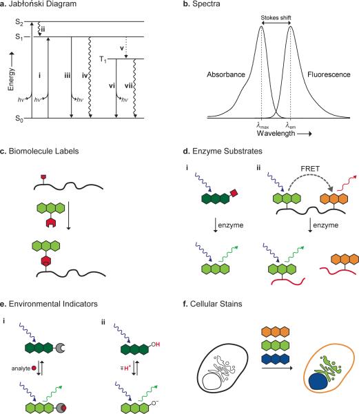
Photophysical concepts (a, b) and biological applications (c–f) of small-molecule fluorophores. a) Jabłoński diagram. i) Absorption of a photon gives an excited state. ii) Internal conversion to S1. iii) Fluorescence. iv) Non-radiative decay. v) Intersystem crossing to T1. vi) Phosphorescence. vii) Non-radiative decay. b) Generic absorption and emission spectra. c) Site-specific labeling of a biomolecule by an orthogonal reaction between two functional groups (red). d) Enzyme substrates. i) Enzyme-catalyzed removal of a blocking group (red) elicits a change in fluorescence. ii) Enzyme catalyzes the cleavage of a labeled biomolecule (red) and concomitant decrease in FRET. e) Environmental indicators. i) Binding of an analyte (red) elicits a change in fluorescence. ii) Protonation of a fluorophore elicits a change in fluorescence. f) Staining of subcellular domains by distinct fluorophores.
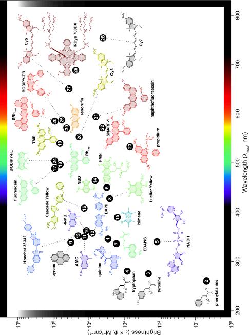
Plot of fluorophore brightness (ε × Φ) versus the wavelength of maximum absorption (λmax) for the major classes of fluorophores. The color of the structure indicates its wavelength of maximum emission (λem). For clarity, only the fluorophoric moiety of some molecules is shown.
References
-
- Petit J-M, Denis-Gay M, Ratinaud M-H. Assessment of fluorochromes for cellular structure and function studies by flow cytometry. Biol. Cell. 1993;78:1–13. - PubMed
-
- Waggoner A, Kenneth S. Covalent labeling of proteins and nucleic acids with fluorophores. Methods Enzymol. 1995;246:362–373. - PubMed
-
- Johnson I. Fluorescent probes for living cells. Histochem. J. 1998;30:123–140. - PubMed
-
- Boonacker E, Van Noorden CJF. Enzyme cytochemical techniques for metabolic mapping in living cells, with special reference to proteolysis. J. Histochem. Cytochem. 2001;49:1473–1486. - PubMed
-
- Zhang J, Campbell RE, Ting AY, Tsien RY. Creating new fluorescent probes for cell biology. Nat. Rev. Mol. Cell Biol. 2002;3:906–918. - PubMed
Publication types
MeSH terms
Substances
Grants and funding
LinkOut - more resources
Full Text Sources
Other Literature Sources
Molecular Biology Databases
