. 2008 Sep;125(1):21-7.
doi: 10.1111/j.1365-2567.2008.02812.x. Epub 2008 Mar 19.

Serum-derived exosomes from antigen-fed mice prevent allergic sensitization in a model of allergic asthma

Affiliations

Serum-derived exosomes from antigen-fed mice prevent allergic sensitization in a model of allergic asthma

Nina Almqvist et al. Immunology. 2008 Sep.

Abstract

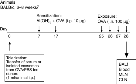
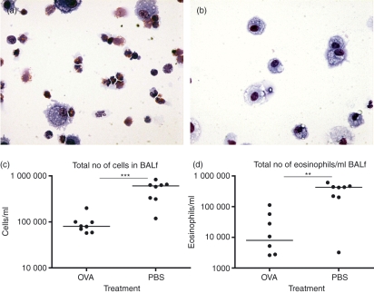
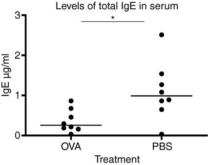
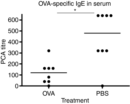
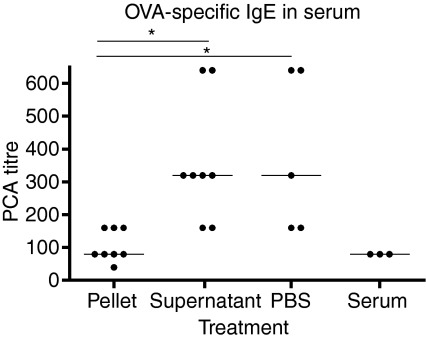
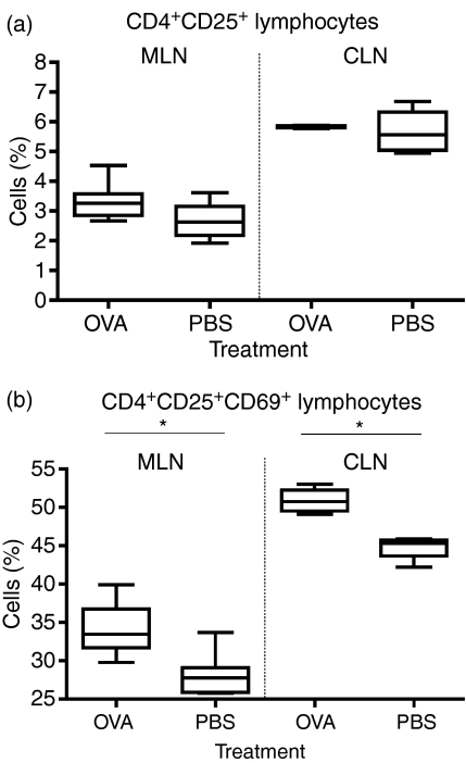
Oral tolerance is an active process that starts with sampling of luminal antigens by the intestinal epithelial cells (IEC), followed by processing and assembly with major histocompatibility complex class II and subsequently a release of tolerogenic exosomes (tolerosomes) from the IEC. We have previously shown that tolerosomes can be isolated from serum shortly after an antigen feed, and will potently transfer antigen-specific tolerance to naive recipients. Here we study the capacity of the tolerosomes to protect against allergic sensitization in a mouse model of allergic asthma. Serum or isolated serum exosomes from tolerized BALB/c donor mice were transferred to syngeneic recipients followed by sensitization and intranasal exposure to ovalbumin (OVA). Blood, bronchoalveolar lavage (BAL) and lymph nodes were sampled 24 hr after the final exposure. The number of eosinophils was counted in BAL fluid and the levels of immunoglobulin E (IgE) and OVA-specific IgE were measured in serum. Mediastinal and coeliac lymph nodes were analysed by flow cytometry. The animals receiving serum from OVA-fed mice displayed significantly lower numbers of airway eosinophils and lower serum levels of total IgE as well as of OVA-specific IgE compared with controls. Moreover, the tolerant animals showed a significantly higher frequency of activated T cells with a regulatory phenotype in both mediastinal and coeliac lymph nodes. The results show that serum or isolated serum exosomes obtained from OVA-fed mice and administered intraperitoneally to naive recipient mice abrogated allergic sensitization in the recipients.


Figures

Figure 1
Figure 1
Overview of the protocol. The serum transfer followed by the allergic airway sensitization protocol. BALf, bronchoalveolar lavage fluid; CLN, coeliac lymph nodes; i.p., intraperitoneal; MLN, mediastinal lymph nodes; OVA, ovalbumin; PBS, phosphate-buffered saline.
Figure 2
Figure 2
Serum from donors fed ovalbumin (OVA) or phosphate-buffered saline (PBS) was transferred to syngeneic recipient mice (BALB/c to BALB/c) followed by allergic sensitization of the recipient. Bronchoalveolar lavage was performed on the animals and cells from the bronchoalveolar lavage fluid (BALf) were counted, followed by staining with May–Grünwald–Giemsa to distinguish the eosinophils. (a) Microscopic image of cells in BALf after allergen exposure from animals transferred with serum isolated from PBS-fed donors and (b) animals receiving serum from the OVA-fed donors. (c) The total number of cells in the BALf. (d) The number of eosinophils/ml of BALf. All data are from one representative experiment out of three.
Figure 3
Figure 3
Total immunoglobulin E (IgE) levels in serum from mice receiving serum from donors fed either ovalbumin (OVA) or phosphate-buffered saline (PBS) followed by allergic sensitization of the recipient. The animals receiving serum from OVA-fed (OVA) donors had significantly lower levels of IgE in serum compared to PBS-fed donors (PBS). The serum levels of IgE in the recipient animals were measured with standard enzyme-linked immunosorbent assay. One representative experiment out of three is shown.
Figure 4
Figure 4
Ovalbumin (OVA) -specific immunoglobulin E (IgE) antibody levels in serum from mice receiving serum from donors fed either OVA or phosphate-buffered saline (PBS) followed by allergic sensitization of the recipient. OVA-specific IgE was measured with passive cutaneus anaphylaxis (PCA) in rats. One representative experiment out of three is shown.
Figure 5
Figure 5
Exosomes were isolated from donor animal serum after feeding them ovalbumin (OVA). Recipient animals were given pellet or serum supernatant, phosphate-buffered saline (PBS) and whole serum as controls. The OVA-specific immunoglobulin E (IgE) in serum was measured with passive cutaneous anaphylaxis (PCA). One representative experiment out of three is shown.
Figure 6
Figure 6
Analysis of T cells in MLN and CLN after serum transfer followed by allergic sensitization. The total CD4+ CD25+ T cell population (a). Analysis of the CD4+ CD25+ T cell population and the expression of the activation marker CD69 (b, P = 0·0019 and P = 0·0286). One representative experiment out of three.

References

    1. Challacombe SJ, Tomasi TB., Jr Systemic tolerance and secretory immunity after oral immunization. J Exp Med. 1980;152:1459–72. - PMC - PubMed
    1. Dahlman-Hoglund A, Dahlgren U, Ahlstedt S, Hanson LA, Telemo E. Bystander suppression of the immune response to human serum albumin in rats fed ovalbumin. Immunology. 1995;86:128–33. - PMC - PubMed
    1. Hultkrantz S, Ostman S, Telemo E. Induction of antigen-specific regulatory T cells in the liver-draining celiac lymph node following oral antigen administration. Immunology. 2005;116:362–72. - PMC - PubMed
    1. Lundin BS, Karlsson MR, Svensson LA, Hanson LA, Dahlgren UI, Telemo E. Active suppression in orally tolerized rats coincides with in situ transforming growth factor-beta (TGF-beta) expression in the draining lymph nodes. Clin Exp Immunol. 1999;116:181–7. - PMC - PubMed
    1. Mowat AM. Anatomical basis of tolerance and immunity to intestinal antigens. Nat Rev Immunol. 2003;3:331–41. - PubMed

Publication types

MeSH terms