

Stereological estimates of dopaminergic, GABAergic and glutamatergic neurons in the ventral tegmental area, substantia nigra and retrorubral field in the rat
- PMID: 18355970
- PMCID: PMC2575227
- DOI: 10.1016/j.neuroscience.2008.01.046
Stereological estimates of dopaminergic, GABAergic and glutamatergic neurons in the ventral tegmental area, substantia nigra and retrorubral field in the rat
Abstract
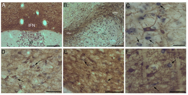
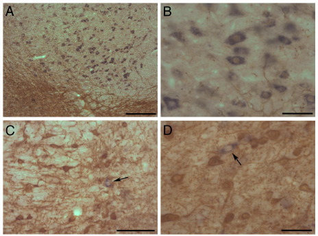
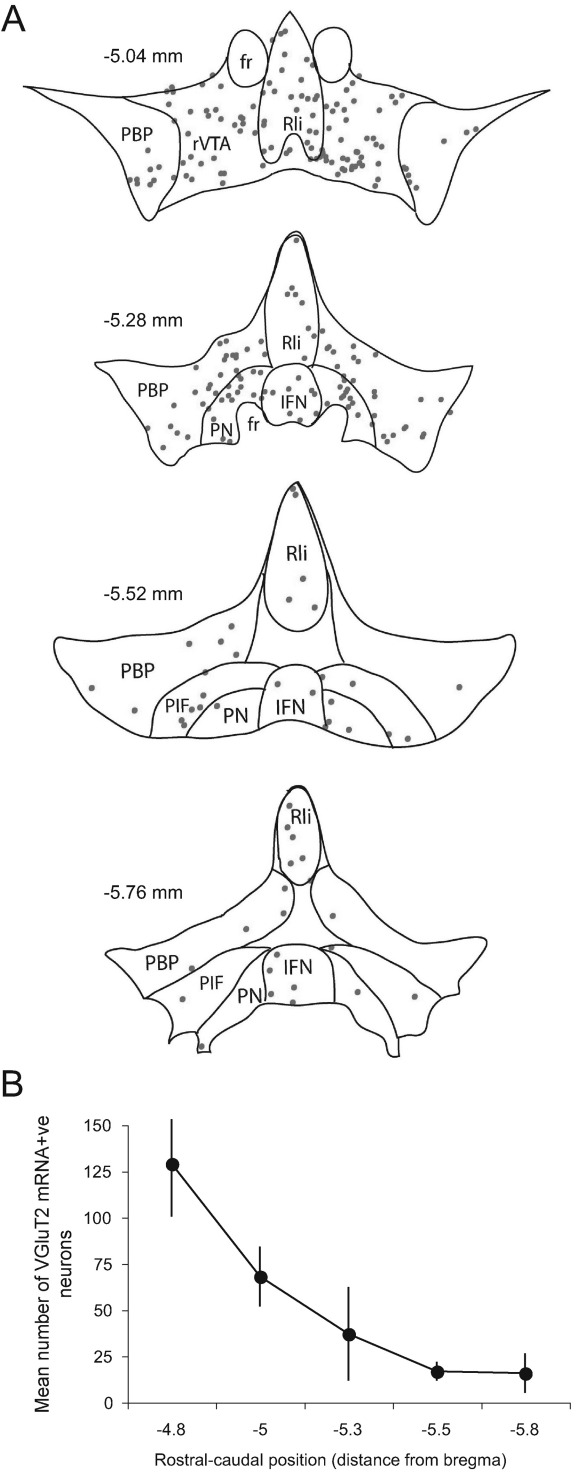
Midbrain dopamine neurons in the ventral tegmental area, substantia nigra and retrorubral field play key roles in reward processing, learning and memory, and movement. Within these midbrain regions and admixed with the dopamine neurons, are also substantial populations of GABAergic neurons that regulate dopamine neuron activity and have projection targets similar to those of dopamine neurons. Additionally, there is a small group of putative glutamatergic neurons within the ventral tegmental area whose function remains unclear. Although dopamine neurons have been intensively studied and quantified, there is little quantitative information regarding the GABAergic and glutamatergic neurons. We therefore used unbiased stereological methods to estimate the number of dopaminergic, GABAergic and glutamatergic cells in these regions in the rat. Neurons were identified using a combination of immunohistochemistry (tyrosine hydroxylase) and in situ hybridization (glutamic acid decarboxylase mRNA and vesicular glutamate transporter 2 mRNA). In substantia nigra pars compacta 29% of cells were glutamic acid decarboxylase mRNA-positive, 58% in the retrorubral field and 35% in the ventral tegmental area. There were further differences in the relative sizes of the GABAergic populations in subnuclei of the ventral tegmental area. Thus, glutamic acid decarboxylase mRNA-positive neurons represented 12% of cells in the interfascicular nucleus, 30% in the parabrachial nucleus, and 45% in the parainterfascicular nucleus. Vesicular glutamate transporter 2 mRNA-positive neurons were present in the ventral tegmental area, but not substantia nigra or retrorubral field. They were mainly confined to the rostro-medial region of the ventral tegmental area, and represented approximately 2-3% of the total neurons counted ( approximately 1600 cells). These results demonstrate that GABAergic and glutamatergic neurons represent large proportions of the neurons in what are traditionally considered as dopamine nuclei and that there are considerable heterogeneities in the proportions of cell types in the different dopaminergic midbrain regions.
Figures
References
-
- Albéri L., Sgadò P., Simon H.H. Engrailed genes are cell-autonomously required to prevent apoptosis in mesencephalic dopaminergic neurons. Development. 2004;131:3229–3236. - PubMed
-
- Albin R.L., Young A.B., Penney J.B. The functional anatomy of basal ganglia disorders. Trends Neurosci. 1989;12:366–375. - PubMed
-
- Bayer V.E., Pickel V.M. GABA-labeled terminals form proportionally more synapses with dopaminergic neurons containing low densities of tyrosine hydroxylase-immunoreactivity in rat ventral tegmental area. Brain Res. 1991;559:44–55. - PubMed
-
- Cameron D.L., Wessendorf M.W., Williams J.T. A subset of ventral tegmental area neurons is inhibited by dopamine, 5-hydroxytryptamine and opioids. Neuroscience. 1997;77:155–166. - PubMed
Publication types
MeSH terms
Substances
Grants and funding
LinkOut - more resources
Full Text Sources
Other Literature Sources
Miscellaneous
