. 2008 May 16;283(20):13707-13.
doi: 10.1074/jbc.M710030200. Epub 2008 Mar 20.

Phosphorylation of MDMX mediated by Akt leads to stabilization and induces 14-3-3 binding

Affiliations

Phosphorylation of MDMX mediated by Akt leads to stabilization and induces 14-3-3 binding

Vanessa Lopez-Pajares et al. J Biol Chem. .

Abstract

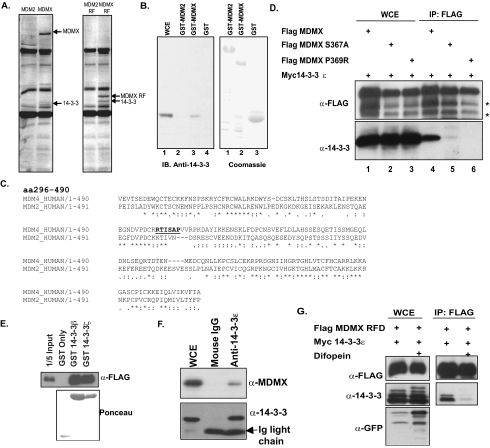
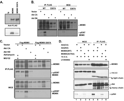
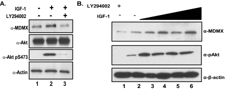
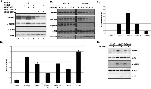
The critical tumor suppressor p53 is mutated or functionally inactivated in nearly all cancers. We have shown previously that the MDM2-MDMX complex functions as an integral unit in targeting p53 for degradation. Here we identify the small protein 14-3-3 as a binding partner of MDMX, which binds at the C terminus (Ser367) in a phosphorylation-dependent manner. Importantly, we demonstrate that the serine/threonine kinase Akt mediates phosphorylation of MDMX at Ser367. This phosphorylation leads to stabilization of MDMX and consequent stabilization of MDM2. Previous studies have shown that Akt phosphorylates and stabilizes MDM2. Our data suggest that stabilization of MDMX by Akt may be an alternative mechanism by which Akt up-regulates MDM2 protein levels and exerts its oncogenic effects on p53 in tumor cells.


Figures

FIGURE 1.
FIGURE 1.
14-3-3 is an MDMX binding partner. A, 293-TREx cells were induced with tetracycline to express FLAG-tagged full-length MDM2 or MDMX or the MDM2 RFD or MDMX RFD. Immunoprecipitation was carried out followed by SDS-PAGE, and unique bands were sent for mass spectrometry. B, GST-MDM2 and GST-MDMX were incubated with lysate expressing 14-3-3ε to determine binding in vitro. WCE, whole cell extract; IB, immunoblotting. C, the sequence alignment of MDM2 and MDMX is shown. The underlined portion indicates the 14-3-3 binding motif. aa, amino acids. D, FLAG-tagged MDMX, MDMX(S367A), and MDMX(P369R) were coexpressed with 14-3-3 in 293T cells. Immunoprecipitation (IP) was carried out against FLAG-tagged proteins using M2 beads. Asterisks indicate degradation products often seen with MDMX overexpression. E, FLAG-tagged MDMX RFD was expressed in 293T cells, and lysates were incubated with GST beads, GST-14-3-3β, or GST-14-3-3ζ. F, Myc-14-3-3ε was coexpressed with FLAG-MDMX in 293T cells. Immunoprecipitation was carried out against Myc-14-3-3ε. G, FLAG-tagged MDMX RFD was coexpressed with 14-3-3ε and with or without difopein in 293T cells. FLAG immunoprecipitation was then carried out, followed by Western blot analysis with the indicated antibodies. GFP, green fluorescent protein.
FIGURE 2.
FIGURE 2.
Akt-mediated phosphorylation of MDMX. A, an in vitro kinase assay was carried out with immunopurified FLAG-tagged MDMX or MDMX(S367A) incubated with cell lysates expressing constitutively active Akt. B, 293T cells were cotransfected with CA-Akt or DN-Akt and FLAG-tagged MDMX or MDMX(S367A). FLAG immunoprecipitation (IP; left panel), whole cell extract analysis (WCE; right panel), and Western blot analysis were carried out using a phospho-specific antibody against Ser367. WT, wild-type. C, 293T cells were cotransfected and analyzed as in B with an additional sample treated with Adriamycin and the proteasome inhibitor MG132. Top panels, immunoprecipitation; bottom panels, whole cell extract. D, cotransfection of 293T cells and immunoprecipitation were carried out as in B with additional treatment with LY294002, and Western blot analysis was carried out with the indicated antibodies. Left, whole cell extract; right, immunoprecipitation.
FIGURE 3.
FIGURE 3.
Endogenous stabilization of MDMX mediated by Akt. A, MCF7 cells were serum-starved overnight and stimulated with IGF-1 for 4 h or pretreated with LY294002 for 30 min and then stimulated with IGF-1. B, MCF7 cells were serum-starved overnight and treated with increasing amounts of IGF-1 (1–50 ng/ml) for 4 h.
FIGURE 4.
FIGURE 4.
Stabilization of MDMX mediated by Akt leads to MDM2 stabilization and p53 down-regulation. A, 293T cells were cotransfected with CA-Akt or KD-Akt and MDMX, MDMX(S367A), or MDMX(C463A). Western blot analysis was carried out with the indicated antibodies. WT, wild-type. B, 293T cells were cotransfected with MDMX, MDM2, and CA-Akt or KD-Akt. Cells were then treated with cycloheximide for the indicated time points. Western blot analysis was carried out with the indicated antibodies. C, a Gal4-Luc mammalian two-hybrid reporter system expressing Gal4-MDM2 and VP16-MDMX alone or with CA-Akt or DN-Akt or expressing MDMX RFD was used in 293T cells. D, H1299 cells were used for a luciferase assay to determine p53 transcriptional activity with the pG13 reporter and p53 alone or with MDMX, CA-Akt, KD-Akt, or a combination of MDMX and Akt. E, U2OS, LNCaP, and OVCA420 cells were treated with the PI 3-kinase inhibitor LY294002, and Western blot analysis was carried out with the indicated antibodies.

References

    1. Vogelstein, B., Lane, D., and Levine, A. J. (2000) Nature 408 307–310 - PubMed
    1. Michael, D., and Oren, M. (2003) Semin. Cancer Biol. 13 49–58 - PubMed
    1. Finch, R. A., Donoviel, D. B., Potter, D., Shi, M., Fan, A., Freed, D. D., Wang, C. Y., Zambrowicz, B. P., Ramirez-Solis, R., Sands, A. T., and Zhang, N. (2002) Cancer Res. 62 3221–3225 - PubMed
    1. Parant, J., Chavez-Reyes, A., Little, N. A., Yan, W., Reinke, V., Jochemsen, A. G., and Lozano, G. (2001) Nat. Genet. 29 92–95 - PubMed
    1. Jackson, M. W., and Berberich, S. J. (2000) Mol. Cell. Biol. 20 1001–1007 - PMC - PubMed

Publication types

MeSH terms