

Case Reports
doi: 10.3174/ajnr.A1038.
Epub 2008 Mar 20.
Prenatal and neonatal MR imaging findings in oral-facial-digital syndrome type VI
Affiliations
- PMID: 18356465
- PMCID: PMC8118817
- DOI: 10.3174/ajnr.A1038
Item in Clipboard
Case Reports
Prenatal and neonatal MR imaging findings in oral-facial-digital syndrome type VI
AJNR Am J Neuroradiol.
2008 Jun.
Abstract
We report prenatal and neonatal neuroimaging findings in a case of oral-facial-digital syndrome type VI (OFDS VI). Prenatal MR imaging at 29 weeks' gestation showed hypoplastic cerebellar vermis and hemispheres, the molar tooth sign, and a hypothalamic hamartoma. Neonatal MR imaging confirmed these findings. The neonate developed breathing abnormalities and exhibited frontal bossing, multiple bucco-alveolar frenula, and postaxial hexadactyly of both hands. If the molar tooth sign and a hypothalamic hamartoma are present, prenatal diagnosis of OFDS VI is possible.
Figures
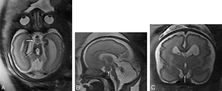
Fetal T2-weighted MR imaging at 29 gestational weeks. A, Axial MR image showing hypoplasia of both cerebellar hemispheres, the characteristic MTS (thin arrow) with thickened and elongated superior cerebellar peduncles and an abnormally deep interpeduncular fossa, and an HH (thick arrow). B, Sagittal MR image revealing significant hypoplasia of the cerebellar vermis (arrow), an enlarged fourth ventricle and posterior fossa, an HH (white arrow), and a thin brain stem. C, Coronal MR image demonstrating an HH (white arrow).
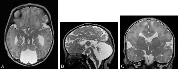
T2-weighted MR imaging at 2 days of age. A, Axial MR image showing hypoplasia of the vermis and both cerebellar hemispheres, the characteristic MTS (thin arrow) with thickened and elongated superior cerebellar peduncles, an abnormally deep interpeduncular fossa, and an HH (thick arrow). B, Sagittal MR image revealing significant hypoplasia of the cerebellar vermis and dysplasia of the remnants of the cerebellar vermis (arrow), an enlarged fourth ventricle and posterior fossa, and an HH (white arrow). In addition, the corpus callosum and the brain stem are hypoplastic, the pituitary stalk is thick, and the interthalamic adhesion is large. C, Coronal MR image demonstrating an HH (arrow), a missing left leaf of the septum pellucidum, a bulky left fornix, and a probably dysplastic cerebral cortex in the left Sylvian fissure.
References
-
- Munke M, McDonald DM, Cronister A, et al. Oral-facial-digital syndrome type VI (Váradi syndrome): further clinical delineation. Am J Med Genet 1990;35:360–69 - PubMed
-
- Parisi MA, Doherty D, Chance PF, et al. Joubert syndrome (and related disorders) (OMIM 213300). Eur J Hum Genet 2007;15:511–21 - PubMed
-
- Maria BL, Boltshauser E, Palmer SC, et al. Clinical features and revised diagnostic criteria in Joubert syndrome. J Child Neurol 1999;14:583–90 - PubMed
-
- Braddock SR, Henley KM, Maria BL. The face of Joubert syndrome: a study of dysmorphology and anthropometry. Am J Med Genet A 2007;143:3235–42 - PubMed
Publication types
MeSH terms
LinkOut - more resources
Full Text Sources
Medical