

Diffusion-weighted MR imaging: diagnosing atypical or malignant meningiomas and detecting tumor dedifferentiation
- PMID: 18356472
- PMCID: PMC8118818
- DOI: 10.3174/ajnr.A0996
Diffusion-weighted MR imaging: diagnosing atypical or malignant meningiomas and detecting tumor dedifferentiation
Abstract
Background and purpose: Atypical and malignant meningiomas are uncommon tumors with aggressive behavior and higher mortality, morbidity, and recurrence compared with benign tumors. We investigated the utility of diffusion-weighted (DW) MR imaging to differentiate atypical/malignant from benign meningiomas and to detect histologic dedifferentiation to higher tumor grade.
Materials and methods: We retrospectively compared conventional and DW MR images (b-value 1000 s/mm(2)) acquired on a 1.5T clinical scanner between 25 atypical/malignant and 23 benign meningiomas. The optimal cutoff for the absolute apparent diffusion coefficient (ADC) and normalized ADC (NADC) ratio to differentiate between the groups was determined by using receiver operating characteristic (ROC) analysis.
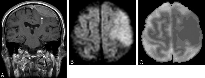
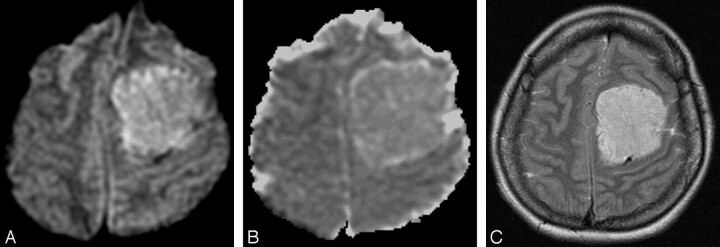
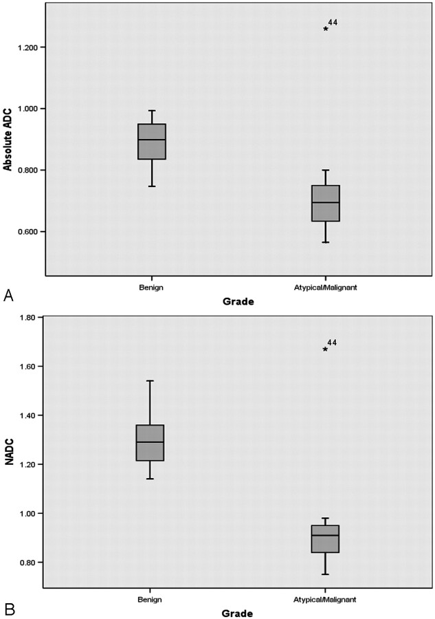
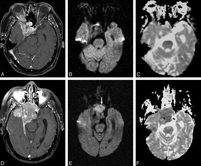
Results: Irregular tumor margins, peritumoral edema, and adjacent bone destruction occurred significantly more often in atypical/malignant than in benign meningiomas. The mean ADC of atypical/malignant meningiomas (0.66 +/- 0.13 x 10(-3) mm(2)/s) was significantly lower compared with benign meningiomas (0.88 +/- 0.08 x 10(-3) mm(2)/s; P < .0001). Mean NADC ratio in the atypical/malignant group (0.91 +/- 0.18) was also significantly lower than the benign group (1.28 +/- 0.11; P < .0001), without overlap between groups. ROC analysis showed that ADC and NADC thresholds of 0.80 x 10(-3) mm(2)/s and 0.99, respectively, had the best accuracy: at the NADC threshold of 0.99, the sensitivity and specificity were 96% and 100%, respectively. Two patients had isointense benign tumors on initial DW MR imaging, and these became hyperintense with the decrease in ADC and NADC below these thresholds when they progressed to atypical and malignant meningiomas on recurrence.
Conclusions: ADC and NADC ratios in atypical/malignant meningiomas are significantly lower than in benign tumors. Decrease in ADC and NADC on follow-up imaging may suggest dedifferentiation to higher tumor grade.
Figures
Comment in
-
Technetium Tc99m-tetrofosmin brain single-photon emission CT for the diagnosis of malignant meningiomas.AJNR Am J Neuroradiol. 2009 Feb;30(2):E21. doi: 10.3174/ajnr.A1286. Epub 2009 Feb 4. AJNR Am J Neuroradiol. 2009. PMID: 19193747 Free PMC article. No abstract available.
References
-
- Das A, Tang WY, Smith DR. Meningiomas in Singapore: demographic and biological characteristics. J Neurooncol 2000;47:153–60 - PubMed
-
- Hakyemez B, Yildirim N, Gokalp G, et al. The contribution of diffusion-weighted MR imaging to distinguishing typical from atypical meningiomas. Neuroradiology 2006;48:513–20 - PubMed
-
- Carpeggiani P, Crisi G, Trevisan C. MRI of intracranial meningiomas: correlations with histology and physical consistency. Neuroradiology 1993;35:532–36 - PubMed
-
- Jaaskelainen J, Haltia M, Laasonen E, et al. The growth rate of intracranial meningiomas and its relation to histology: an analysis of 43 patients. Surg Neurol 1985;24:165–72 - PubMed
-
- Mahmood A, Caccamo DV, Tomecek FJ, et al. Atypical and malignant meningiomas: a clinicopathological review. Neurosurgery 1993;33:955–63 - PubMed
Publication types
MeSH terms
LinkOut - more resources
Full Text Sources