. 2008 May 9;102(9):1082-90.
doi: 10.1161/CIRCRESAHA.107.167072. Epub 2008 Mar 20.

Inhibition of mitochondrial permeability transition pore opening by ischemic preconditioning is probably mediated by reduction of oxidative stress rather than mitochondrial protein phosphorylation

Affiliations

Inhibition of mitochondrial permeability transition pore opening by ischemic preconditioning is probably mediated by reduction of oxidative stress rather than mitochondrial protein phosphorylation

Samantha J Clarke et al. Circ Res. .

Abstract

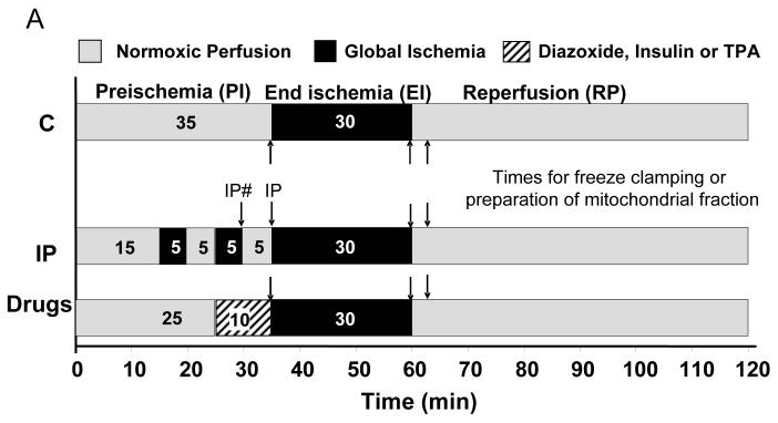
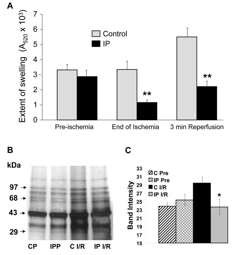
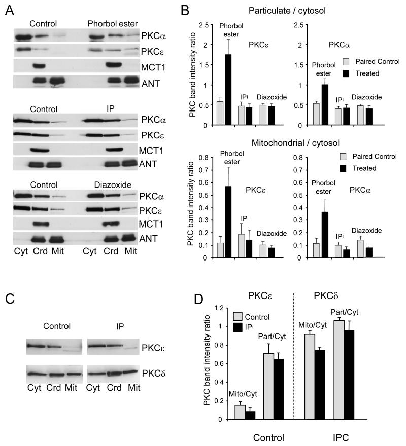
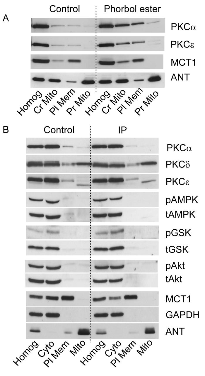
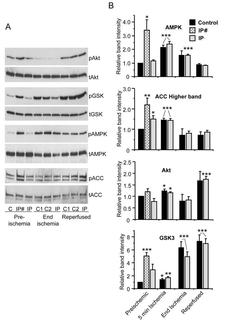
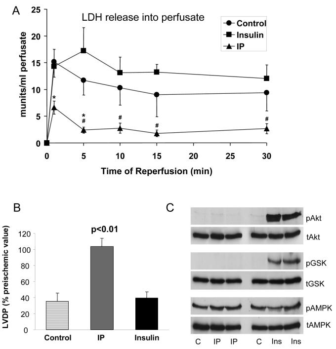
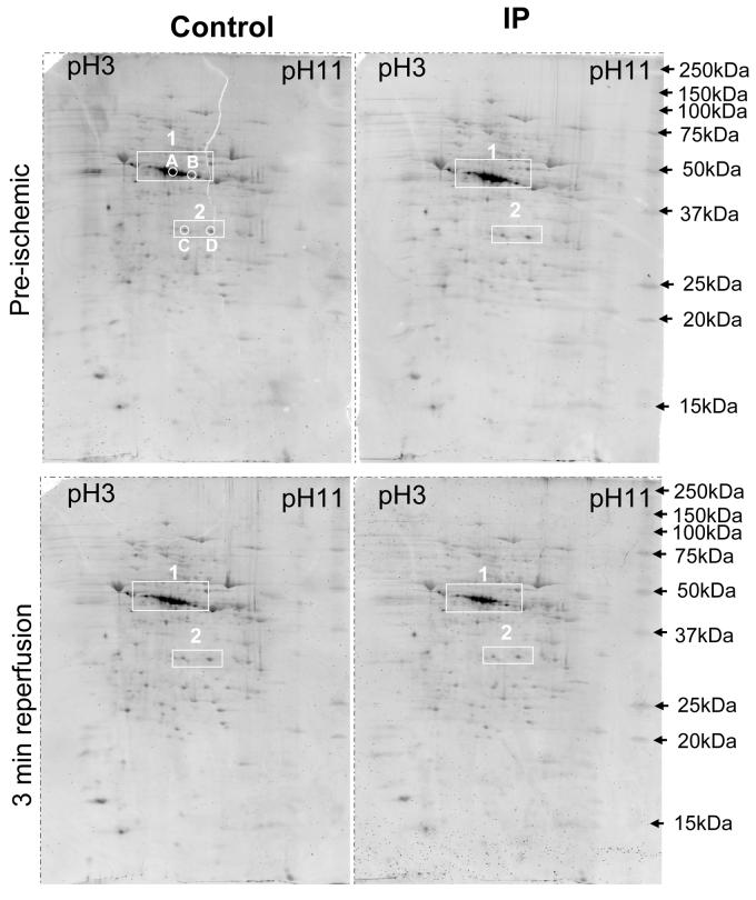
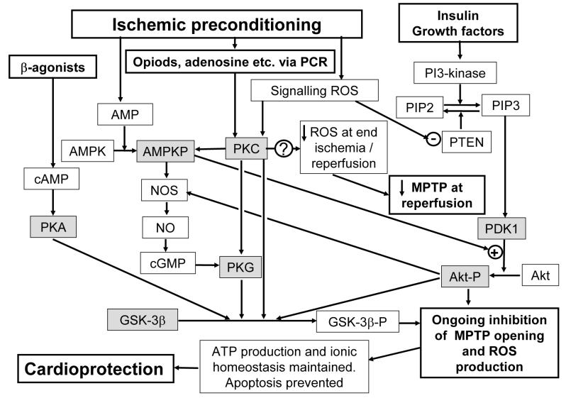
Inhibition of mitochondrial permeability transition pore (MPTP) opening at reperfusion is critical for cardioprotection by ischemic preconditioning (IP). Some studies have implicated mitochondrial protein phosphorylation in this effect. Here we confirm that mitochondria rapidly isolated from preischemic control and IP hearts show no significant difference in calcium-mediated MPTP opening, whereas IP inhibits MPTP opening in mitochondria isolated from IP hearts following 30 minutes of global normothermic ischemia or 3 minutes of reperfusion. Analysis of protein phosphorylation in density-gradient purified mitochondria was performed using both 2D and 1D electrophoresis, with detection of phosphoproteins using Pro-Q Diamond or phospho-amino-specific antibodies. Several phosphoproteins were detected, including voltage-dependent anion channels isoforms 1 and 2, but none showed significant IP-mediated changes either before ischemia or during ischemia and reperfusion, and neither Western blotting nor 2D fluorescence difference gel electrophoresis detected translocation of protein kinase C (alpha, epsilon, or delta isoforms), glycogen synthase kinase 3beta, or Akt to the mitochondria following IP. In freeze-clamped hearts, changes in phosphorylation of GSK3beta, Akt, and AMP-activated protein kinase were detected following ischemia and reperfusion but no IP-mediated changes correlated with MPTP inhibition or cardioprotection. However, measurement of mitochondrial protein carbonylation, a surrogate marker for oxidative stress, suggested that a reduction in mitochondrial oxidative stress at the end of ischemia and during reperfusion may account for IP-mediated inhibition of MPTP. The signaling pathways mediating this effect and maintaining it during reperfusion are discussed.


Figures

Figure 1
Figure 1. Protocols used for heart perfusion
The times at which control (C) and IP-hearts were freeze clamped or homogenised for mitochondrial preparation are indicated by arrows. For pre-ischemic IP hearts samples were taken at the two points indicated as IP# and IP.
Figure 2
Figure 2. MPTP opening and protein carbonylation in mitochondria from control and IP-hearts
Panel A reports the maximum decrease in A520 after calcium addition to de-energised mitochondria. Panel B shows a representative western blot for protein carbonylation (CP – non-ischemic control hearts). Data in Panels A and C are presented as means of 6 or 12 (preischemic) separate mitochondrial preparations from each group (IP versus control: * p<0.05, ** p<0.01).
Figure 3
Figure 3. IP does not cause translocation of protein kinase C isoforms to mitochondria
Sub-cellular fractionation of hearts into cytosolic (Cyt), crude mitochondria (Crd) and pure mitochondria (Mit) was performed according to protocol 1 (Supplementary Fig. 1S). Panels A and B represent fractions isolated immediately after IP or following 10 min treatment with 50 μmol/L diazoxide or 0.2 μmol/L phorbol ester, whilst Panels C and D represent fractions isolated after 30 min ischemia and 3 min reperfusion. Proteins were separated by SDS-PAGE followed by western blotting with the appropriate antibody. Panels B and D present mean data (n = 6) of the ratio of mitochondrial or crude particulate PKC to cytosolic PKC.
Figure 4
Figure 4. IP does not cause mitochondrial recruitment or phosphorylation of other protein kinases
Sub-cellular fractionation of heart homogenates (Homog) into cytosolic (Cyt), plasma membrane (Pl Mem) and pure mitochondria (Mit) was performed according to protocol 2 (see Supplementary Figure 1S) immediately after the IP protocol (Panel B) or after 10 min with 0.2 μmol/L phorbol ester (Panel A ). Representative western blots with the antibodies indicated are representative of at least 3 experiments. Glyceraldehyde-3-phosphate dehydrogenase (GAPDH) was used as a cytosolic marker.
Figure 5
Figure 5. Cytosolic AMPK, Akt, GSK3β and ACC phosphorylation state in freeze-clamped hearts
Control and IP hearts were freeze-clamped at the times shown in Figure 1 and a cytosolic fraction produced according to the freeze-clamp protocol of Supplementary Figure 1S. Proteins were separated by SDS-PAGE followed by western blotting with the appropriate antibody for the total (t) or phosphorylated (p) kinases indicated or for acetylCoA carboxylase (ACC). Panel A shows representative blots whilst panel B shows mean data (n = 6) of scanned blots where the ratio of phosphorylated to total protein is expressed relative to the ratio for the control preischemic sample run on the same gel. IP# and IP samples represent samples taken from IP hearts at the end of the third brief ischemic phase of preconditioning or immediately before ischemia as indicated in Figure 1. In Panel A, where two adjacent control samples (C) are shown, they represent 2 separate hearts. * p<0.05; ** p<0.02; *** p<0.01 versus preischemic control.
Figure 6
Figure 6. Insulin treatment prior to ischemia increases Akt and GSK3β phosphorylation but is not cardioprotective
Hearts were pre-treated with 0.7 nmol/L insulin for 19 min followed by 1 minute of washout prior to 30 min ischemia and then reperfusion for the time shown. Panel A shows the LDH released into the perfusate during reperfusion whist Panel B shows the recovery of LVDP after 30 min reperfusion as a percentage of the pre-ischemic value. Panel C presents data from hearts that were freeze-clamped prior to ischemia and then treated as described for Figure 5.
Figure 7
Figure 7. IP-mediated changes in mitochondrial protein phosphorylation were not detected
Mitochondria were isolated from control or IP hearts just prior to ischemia (Pre) or following 3 min reperfusion (Rep), separated by 2D-gel electrophoresis and then stained with the phosphoprotein stain, Pro-Q Diamond. In supplementary Figure 2A the same data are shown in red overlaid on the total protein stain (Sypro-Ruby) in green. The data shown in Supplementary Fig. 9s and Table 2 establish the identity of the spots in box 1 as different phosphorylation states of PDHE1α and those in box 2 as singly phosphorylated VDAC1 and VDAC2 .
Figure 8
Figure 8. Suggested pathways by which IP may lead to inhibition of MPTP opening during reperfusion
Active protein kinases are shaded grey. Further details are given in the text.

References

    1. Halestrap AP, Clarke SJ, Javadov SA. Mitochondrial permeability transition pore opening during myocardial reperfusion - a target for cardioprotection. Cardiovasc Res. 2004;61:372–385. - PubMed
    1. Yellon DM, Downey JM. Preconditioning the myocardium: From cellular physiology to clinical cardiology. Physiol Rev. 2003;83:1113–1151. - PubMed
    1. Javadov SA, Clarke S, Das M, Griffiths EJ, Lim KHH, Halestrap AP. Ischaemic preconditioning inhibits opening of mitochondrial permeability transition pores in the reperfused rat heart. J Physiol. 2003;549:513–524. - PMC - PubMed
    1. Argaud L, Gateau-Roesch O, Chalabreysse L, Gomez L, Loufouat J, Thivolet-Bejui F, Robert D, Ovize M. Preconditioning delays Ca2+-induced mitochondrial permeability transition. Cardiovasc Res. 2004;61:115–122. - PubMed
    1. Khaliulin I, Schwalb H, Wang P, Houminer E, Grinberg L, Katzeff H, Borman JB, Powell SR. Preconditioning improves postischemic mitochondrial function and diminishes oxidation of mitochondrial proteins. Free Radic Biol Med. 2004;37:1–9. - PubMed

Publication types

MeSH terms

Substances