

Sympathetic activation causes focal adhesion signaling alteration in early compensated volume overload attributable to isolated mitral regurgitation in the dog
- PMID: 18356543
- PMCID: PMC3092391
- DOI: 10.1161/CIRCRESAHA.107.163642
Sympathetic activation causes focal adhesion signaling alteration in early compensated volume overload attributable to isolated mitral regurgitation in the dog
Expression of concern in
-
Expression of Concern: Sympathetic Activation Causes Focal Adhesion Signaling Alteration in Early Compensated Volume Overload Attributable to Isolated Mitral Regurgitation in the Dog.Circ Res. 2023 Mar 31;132(7):e114. doi: 10.1161/RES.0000000000000604. Epub 2023 Mar 13. Circ Res. 2023. PMID: 36912159 No abstract available.
Abstract
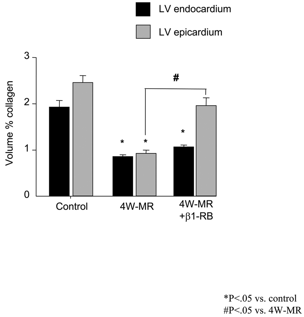
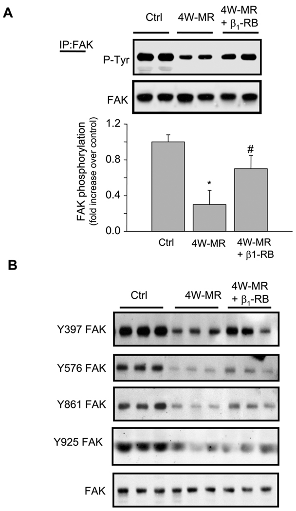
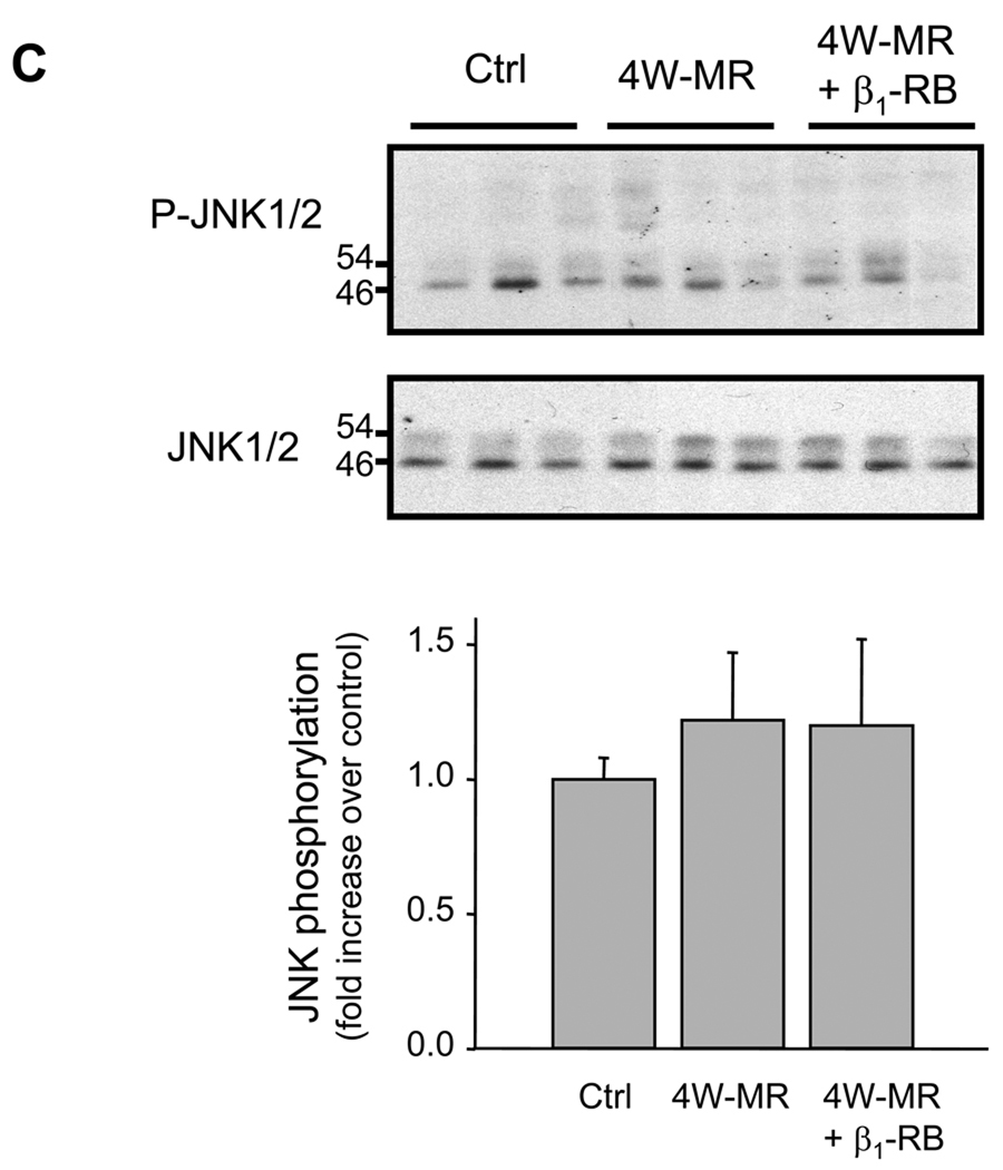
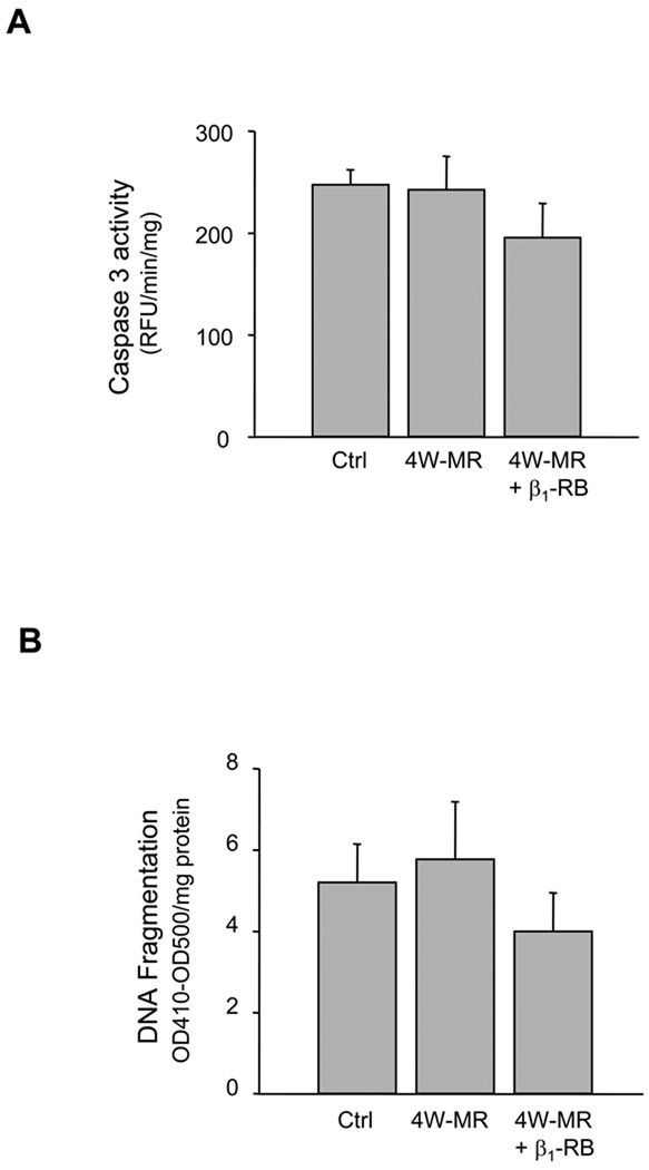
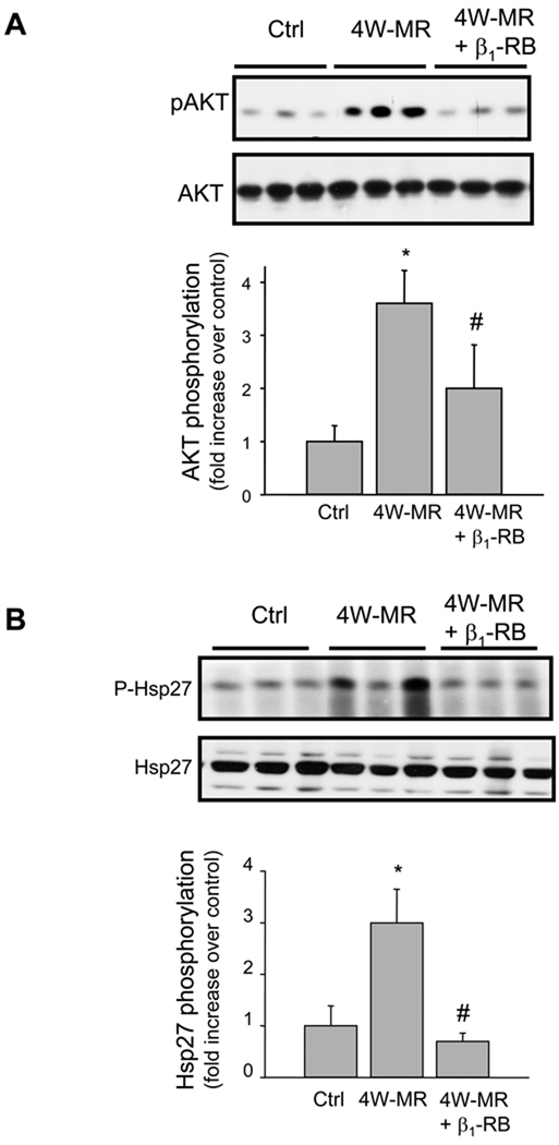
We reported that left ventricular (LV) dilatation after 4 weeks of isolated mitral regurgitation (MR) in the dogs is marked by extracellular matrix loss and an increase in adrenergic drive. Given that extracellular matrix proteins and their receptor integrins influence beta-adrenergic receptor (beta-AR) responses in vitro, we tested whether beta1-AR activation modulates focal adhesion (FA) signaling and LV remodeling in these same dogs with isolated MR. Normal dogs were compared with dogs with MR of a 4-week duration and with MR dogs treated with beta(1)-AR blockade (beta(1)-RB) (extended-release metoprolol succinate, 100 mg QD) that was started 24 hours after MR induction. In MR LVs, a decrease in collagen accumulation compared with normal dogs was associated with a decrease in FA kinase tyrosine phosphorylation, along with FA kinase interaction with adapter and cytoskeletal proteins, p130(Cas) and paxillin, respectively, as determined by immunoprecipitation assays. There was increased phosphorylation of stress related molecules p38 mitogen-activated protein kinase (MAPK) and Hsp27 and survival signaling kinases extracellular signal-regulated kinase 1/2 and AKT, with no evidence of cardiomyocyte apoptosis. beta(1)-RB attenuated FA signaling loss and prevented p38 MAPK, Hsp27, and AKT phosphorylation induced by MR and significantly increased LV epicardial collagen content. However, beta(1)-RB did not improve LV endocardial collagen loss or LV dilatation induced by MR. Isolated myocytes from normal and MR dog hearts treated with beta(1)- or beta(2)-AR agonists demonstrated no difference in FA kinase, p38 MAPK, Hsp27, or AKT phosphorylation. These results showed that chronic stimulation of beta(1)-AR during early compensated MR impairs FA signaling that may affect myocyte/fibroblast-extracellular matrix scaffolding necessary for LV remodeling.
Figures


Similar articles
-
Beta1-adrenergic receptors promote focal adhesion signaling downregulation and myocyte apoptosis in acute volume overload.J Mol Cell Cardiol. 2012 Aug;53(2):240-9. doi: 10.1016/j.yjmcc.2012.05.004. Epub 2012 May 15. J Mol Cell Cardiol. 2012. PMID: 22609523 Free PMC article.
-
Dissociation between cardiomyocyte function and remodeling with beta-adrenergic receptor blockade in isolated canine mitral regurgitation.Am J Physiol Heart Circ Physiol. 2008 Dec;295(6):H2321-7. doi: 10.1152/ajpheart.00746.2008. Epub 2008 Oct 10. Am J Physiol Heart Circ Physiol. 2008. PMID: 18849331 Free PMC article.
-
Beta1-adrenergic receptor blockade attenuates angiotensin II-mediated catecholamine release into the cardiac interstitium in mitral regurgitation.Circulation. 2003 Jul 15;108(2):225-30. doi: 10.1161/01.CIR.0000079226.48637.5A. Epub 2003 Jul 7. Circulation. 2003. PMID: 12847066
-
Paxillin interactions.J Cell Sci. 2000 Dec;113 Pt 23:4139-40. doi: 10.1242/jcs.113.23.4139. J Cell Sci. 2000. PMID: 11069756 Review.
-
Left ventricular remodeling in preclinical experimental mitral regurgitation of dogs.J Vet Cardiol. 2012 Mar;14(1):73-92. doi: 10.1016/j.jvc.2012.01.012. Epub 2012 Mar 2. J Vet Cardiol. 2012. PMID: 22386719 Review.
Cited by
-
Beta1-adrenergic receptors promote focal adhesion signaling downregulation and myocyte apoptosis in acute volume overload.J Mol Cell Cardiol. 2012 Aug;53(2):240-9. doi: 10.1016/j.yjmcc.2012.05.004. Epub 2012 May 15. J Mol Cell Cardiol. 2012. PMID: 22609523 Free PMC article.
-
A Comparison of Phenomenologic Growth Laws for Myocardial Hypertrophy.J Elast. 2017 Dec;129(1-2):257-281. doi: 10.1007/s10659-017-9631-8. Epub 2017 Mar 1. J Elast. 2017. PMID: 29632418 Free PMC article.
-
The multiple mechanistic faces of a pure volume overload: implications for therapy.Am J Med Sci. 2014 Oct;348(4):337-46. doi: 10.1097/MAJ.0000000000000255. Am J Med Sci. 2014. PMID: 24781435 Free PMC article. Review.
-
Mitral regurgitation: has another magic bullet bitten the dust?Circ Heart Fail. 2013 Jul;6(4):624-6. doi: 10.1161/CIRCHEARTFAILURE.113.000409. Circ Heart Fail. 2013. PMID: 23861504 Free PMC article. No abstract available.
-
Integrins and integrin-associated proteins in the cardiac myocyte.Circ Res. 2014 Jan 31;114(3):572-586. doi: 10.1161/CIRCRESAHA.114.301275. Circ Res. 2014. PMID: 24481847 Free PMC article. Review.
References
-
- Tallaj J, Wei C-C, Hankes GH, Holland M, Rynders P, Dillon AR, Ardell JL, Armour JA, Lucchesi PA, Dell'Italia LJ. {beta}1-adrenergic receptor blockade attenuates angiotensin II-mediated catecholamine release into the cardiac interstitium in mitral regurgitation. Circulation. 2003;108:225–230. - PubMed
-
- Stewart J, James A, Wei C-C, Brower GL, Rynders PE, Hankes GH, Dillon AR, Lucchesi PA, Janicki JS, Dell'Italia LJ. Cardiac mast cell- and chymase-mediated matrix metalloproteinase activity and left ventricular remodeling in mitral regurgitation in the dog. J Mol Cell Cardiol. 2003;35:311–319. - PubMed
-
- Dell'italia LJ, Balcells E, Meng QC, Su X, Schultz D, Bishop SP, Machida N, Straeter-Knowlen IM, Hankes GH, Dillon R, Cartee RE, Oparil S. Volume-overload cardiac hypertrophy is unaffected by ACE inhibitor treatment in dogs. Am J Physiol Heart Circ Physiol. 1997;273:H961–H970. - PubMed
-
- Perry GJ, Wei C-C, Hankes GH, Dillon SR, Rynders P, Mukherjee R, Spinale FG, Dell'Italia LJ. Angiotensin II receptor blockade does not improve left ventricular function and remodeling in subacute mitral regurgitation in the dog. J Am Coll Cardiol. 2002;39:1374–1379. - PubMed
Publication types
MeSH terms
Substances
Grants and funding
LinkOut - more resources
Full Text Sources
Research Materials
Miscellaneous
