

Enhanced expression of Lp-PLA2 and lysophosphatidylcholine in symptomatic carotid atherosclerotic plaques
- PMID: 18356547
- PMCID: PMC4360896
- DOI: 10.1161/STROKEAHA.107.503193
Enhanced expression of Lp-PLA2 and lysophosphatidylcholine in symptomatic carotid atherosclerotic plaques
Abstract
Background and purpose: Circulating lipoprotein-associated phospholipase A(2) (Lp-PLA(2)) has emerged as a novel biomarker for cardiovascular diseases. However, the correlation between the plaque expression of Lp-PLA(2) and plaque oxidative stress, inflammation, and stability as well as the clinical presentation remains poorly defined, especially for cerebrovascular disease. Therefore, this study was performed to test the hypothesis that Lp-PLA(2) expression is higher in symptomatic than in asymptomatic carotid plaques of patients undergoing carotid endarterectomy.
Methods: The expression of Lp-PLA(2) in 167 carotid artery plaques was determined by immunoblotting and immunostaining. Plaque oxidative stress, inflammation, and stability were quantified by NAD(P)H oxidase p67phox and MMP-2 immunoblotting, oxidized LDL (oxLDL) immunoreactivity, macrophage and Sirius red collagen staining. Lysophosphatidylcholine 16:0 (lysoPC) concentration was measured in 55 plaques using liquid chromatography tandem mass spectrometry.
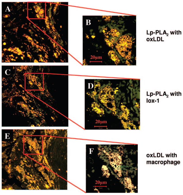
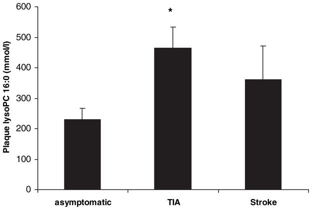
Results: Lp-PLA(2) expression was significantly higher in plaques of symptomatic patients than asymptomatic patients (1.66+/-0.19 versus 1.14+/-0.10, P<0.05) and localized mainly to shoulder and necrotic lipid core areas in colocalization with oxLDL and macrophage content. Similarly, Lp-PLA(2) expression was related to collagen content, which was lower in plaques from symptomatic patients than in plaques from asymptomatic patients (9.1+/-2.2 versus 18.5+/-1.7% of staining/field, P<0.001). LysoPC plaque concentration was significantly higher in plaques of symptomatic than asymptomatic patients (437.0+/-57.91 versus 228.84+/-37.00 mmol/L, P<0.05).
Conclusions: Symptomatic carotid artery plaques are characterized by increased levels of Lp-PLA(2) and its product lysoPC in correlation with markers of tissue oxidative stress, inflammation, and instability. These findings strongly support a role for Lp-PLA2 in the pathophysiology and clinical presentation of cerebrovascular disease.
Figures


References
-
- MacPhee CH, Moores KE, Boyd HF, Dhanak D, Ife RJ, Leach CA, Leake DS, Milliner KJ, Patterson RA, Suckling KE, Tew DG, Hickey DM. Lipoprotein-associated phospholipase a2, platelet-activating factor acetylhydrolase, generates two bioactive products during the oxidation of low-density lipoprotein: Use of a novel inhibitor. Biochem J. 1999;338:479– 487. - PMC - PubMed
-
- Asano KOS, Fukunaga K, Shiomi T, Mori T, Iwata M, Ikeda Y, Yamaguchi K. Cellular source(s) of platelet-activating-factor acetylhydrolase activity in plasma. Biochem Biophys Res Commun. 1999;261:511–514. - PubMed
-
- Stafforini DM, Elstad MR, McIntyre TM, Zimmerman GA, Prescott SM. Human macrophages secret platelet-activating factor acetylhydrolase. J Biol Chem. 1990;265:9682–9687. - PubMed
-
- Hakkinen T, Luoma JS, Hiltunen MO, Macphee CH, Milliner KJ, Patel L, Rice SQ, Tew DG, Karkola K, Yla-Herttuala S. Lipoprotein-associated phospholipase a(2), platelet-activating factor acetylhydrolase, is expressed by macrophages in human and rabbit atherosclerotic lesions. Arterioscler Thromb Vasc Biol. 1999;19:2909–2917. - PubMed
-
- Caslake MJ, Packard CJ, Suckling KE, Holmes SD, Chamberlain P, Macphee CH. Lipoprotein-associated phospholipase a(2), platelet-activating factor acetylhydrolase: A potential new risk factor for coronary artery disease. Atherosclerosis. 2000;150:413– 419. - PubMed
Publication types
MeSH terms
Substances
Grants and funding
LinkOut - more resources
Full Text Sources
Other Literature Sources
Medical
Miscellaneous
