

Three-dimensional mapping of hippocampal anatomy in adolescents with bipolar disorder
- PMID: 18356767
- PMCID: PMC2773145
- DOI: 10.1097/CHI.0b013e31816765ab
Three-dimensional mapping of hippocampal anatomy in adolescents with bipolar disorder
Abstract
Objective: Early-onset bipolar disorder is thought to be a particularly severe variant of the illness. Continuity with the adult form of illness remains unresolved, but preliminary evidence suggests similar biological underpinnings. Recently, we observed localized hippocampal decreases in unmedicated adults with bipolar disorder that were not detectable with conventional volumetric measures. Using the same three-dimensional mapping methods, we sought to investigate whether a similar pattern exists in adolescents with bipolar disorder.
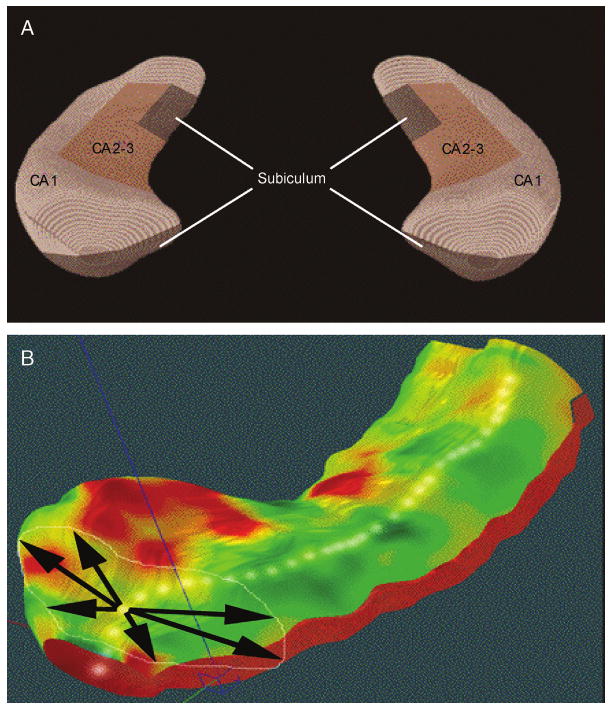
Method: High-resolution brain magnetic resonance images were acquired from 16 adolescents meeting DSM-IV criteria for bipolar disorder (mean age 15.5 +/- 3.4 years, 50% female) and 20 demographically matched, typically developing control subjects. Three-dimensional parametric mesh models of the hippocampus were created from manual tracings of the hippocampal formation.
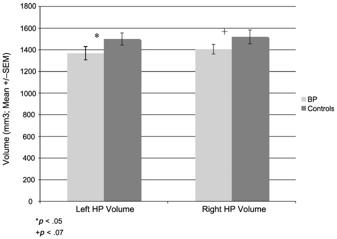
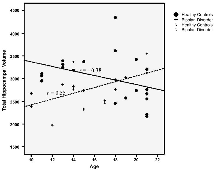
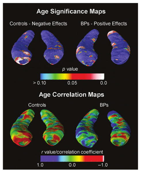
Results: Controlling for total brain volume, total hippocampal volume was significantly smaller in adolescent patients with bipolar disorder relative to controls (by 9.2%). Statistical mapping results, confirmed by permutation testing, revealed significant localized deformations in the head and tail of the left hippocampus in adolescents with bipolar disorder, relative to normal controls. In addition, there was a significant positive correlation between hippocampal size and age in patients with bipolar disorder, whereas healthy controls showed an inverse relation.
Discussion: Localized hippocampal deficits in adolescent patients with bipolar disorder suggest a possible neural correlate for memory deficits observed in this illness. Moreover, age-related increases in hippocampal size in patients with bipolar disorder, not observed in healthy controls, may reflect abnormal developmental mechanisms in bipolar disorder. This possibility must be confirmed by longitudinal studies.
Conflict of interest statement
The other authors report no conflicts of interest.
Figures

Comment in
-
The next wave in neuroimaging research in pediatric bipolar disorder.J Am Acad Child Adolesc Psychiatry. 2008 May;47(5):483-485. doi: 10.1097/CHI.0b013e318168457f. J Am Acad Child Adolesc Psychiatry. 2008. PMID: 18438184 No abstract available.
References
-
- Bannerman DM, Rawlins JN, McHugh SB, et al. Regional dissociations within the hippocampus–memory and anxiety. Neurosci Biobehav Rev. 2004;28:273–283. - PubMed
-
- Bearden CE, Glahn DC, Monkul ES, et al. Sources of declarative memory impairment in bipolar disorder: mnemonic processes and clinical features. J Psychiatr Res. 2006;40:47–58. - PubMed
-
- van Gorp WG, Altshuler L, Theberge DC, Mintz J. Declarative and procedural memory in bipolar disorder. Biol Psychiatry. 1999;46:525–531. - PubMed
-
- Altshuler LL, Ventura J, van Gorp WG, et al. Neurocognitive function in clinically stable men with bipolar I disorder or schizophrenia and normal control subjects. Biol Psychiatry. 2004;56:560–569. - PubMed
