. 2008 May 1;317(1):325-35.
doi: 10.1016/j.ydbio.2008.02.029. Epub 2008 Mar 4.

A novel role for zebrafish zic2a during forebrain development

Affiliations

A novel role for zebrafish zic2a during forebrain development

Nicholas A Sanek et al. Dev Biol. .

Abstract

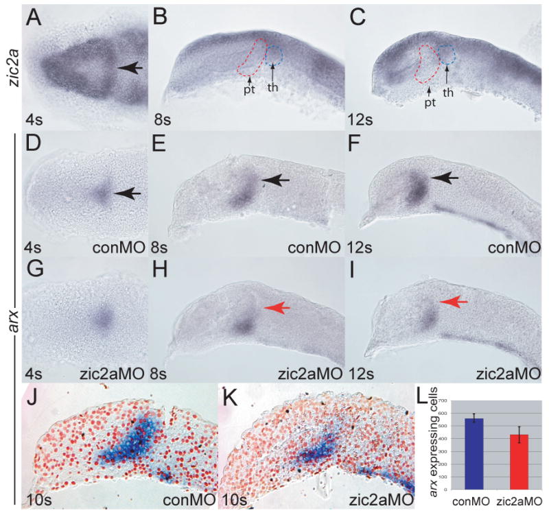
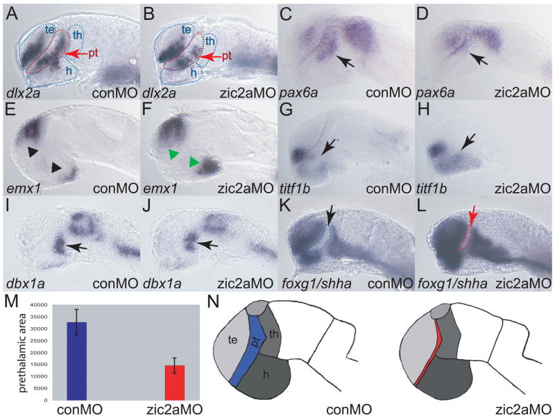
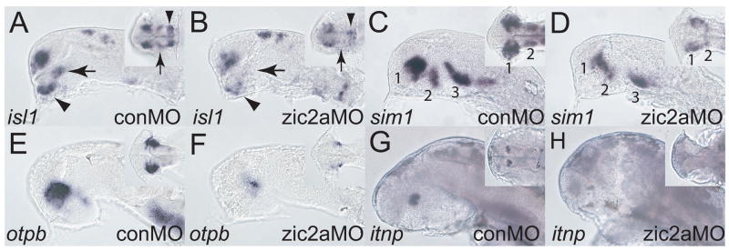
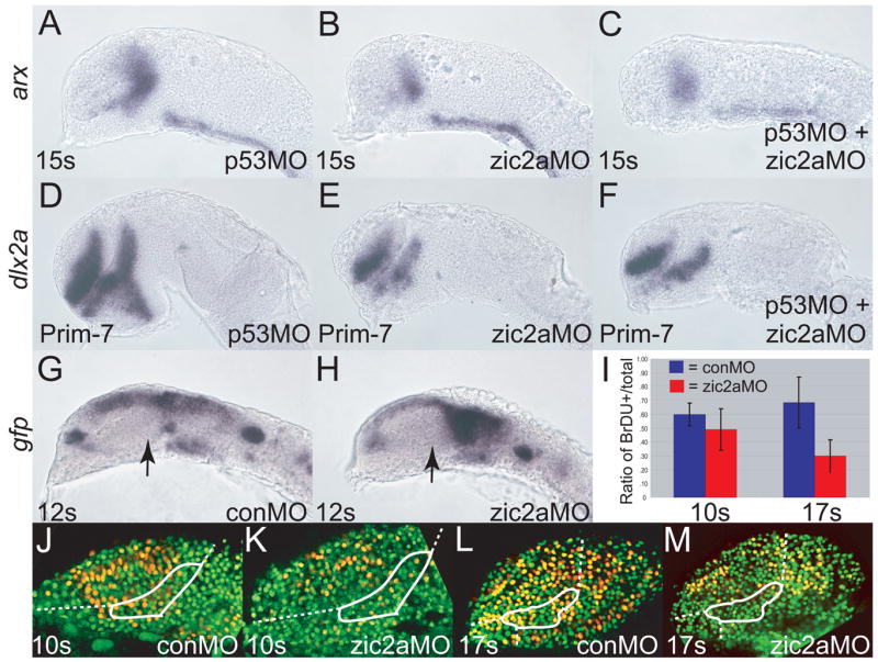
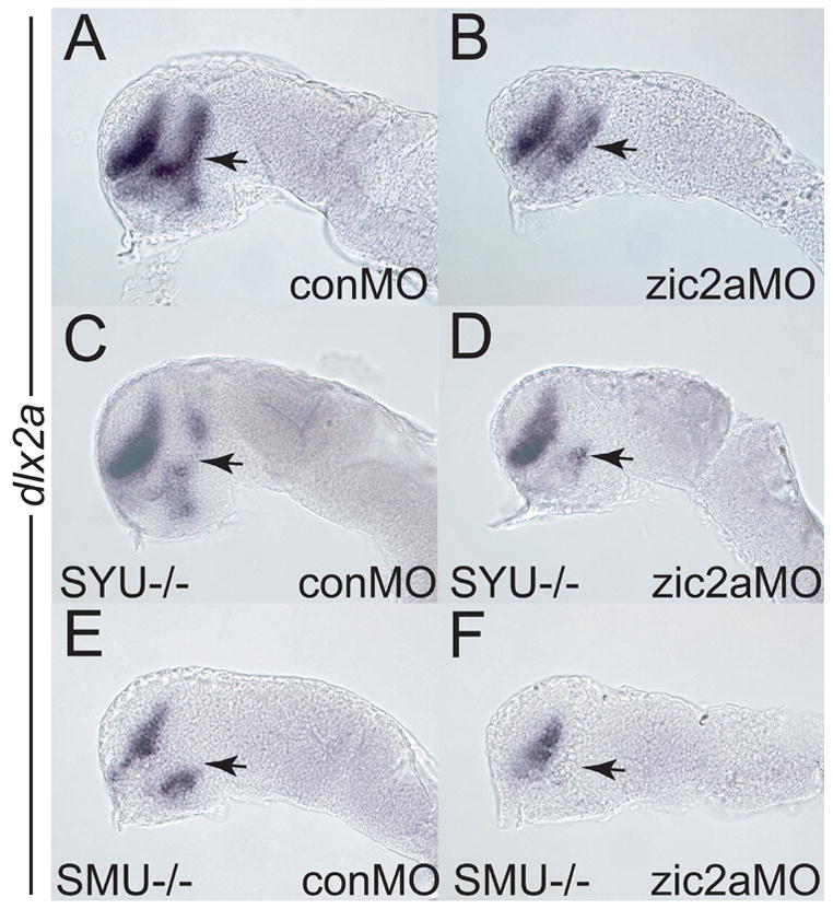
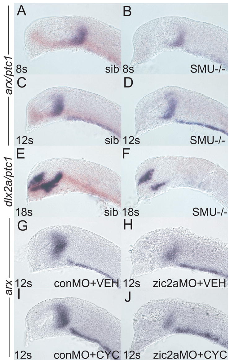
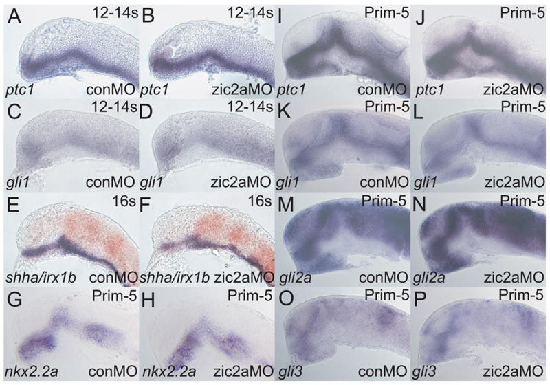
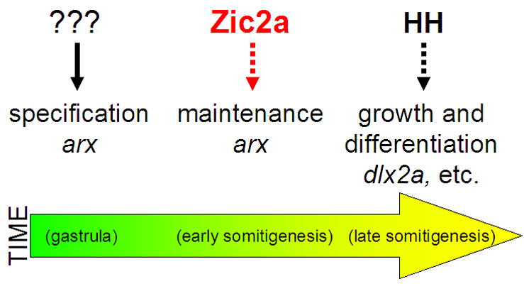
Patterns of transcription factor expression establish a blueprint for the vertebrate forebrain early in embryogenesis. In the future diencephalon, several genes with patterned expression have been identified, yet their specific functions and interactions between them are not well understood. We have uncovered a crucial role for one such gene, zic2a, during formation of the anterior diencephalon in zebrafish. We show that zic2a is required for transcription of the prethalamic markers arx and dlx2a. This function is required during early steps of prethalamic development, soon after its specification. zic genes are evolutionarily related to glis, transcription factors that mediate hedgehog signaling. Intriguingly, the hedgehog signaling pathway also acts to promote development of the prethalamus. We asked if zic2a interacts with hedgehog signaling in the context of forebrain development in zebrafish. Our data show that hedgehog signaling and zic2a function at different times, and therefore act in parallel pathways during forebrain development. Taken together, our results identify Zic2a as a novel regulator of prethalamic development, and show that it functions independently of hedgehog signaling.


Figures

Fig. 1
Fig. 1. Zic2a is required for maintenance, but not for initiation of arx expression in the PT primordium
Embryos were stained by ISH for expression of zic2a (A-C) or arx (D-I). (A) Uninjected embryos express zic2a transiently in the early PT at 4 somites. (B, C) zic2a is not expressed in the PT at 8 and 12 somites. Prethalamus (pt) and thalamus (th) are outlined for reference. (D-E) Normal arx expression in the PT primordium of control MO injected embryos at 4, 8 and 12 somites. (G-H) arx expression in zic2a morphants. (G) arx is expressed normally at 4s (44/50 embryos, 3 exp.). (H) arx is mildly reduced at 8 somites (8/23 embryos, 2 exp.). (I) arx is drastically reduced at 12 somites (28/33 embryos, 2 exp.). (J, K) Representative parasagittal sections of conMO-and zic2aMO-injected embryos stained for arx by ISH at 10s. (L) Graph of average number of arx expressing cells in conMO (n = 3) and zic2aMO-injected (n = 3) embryos (results significant at p = 0.05). All embryos are shown with anterior to the left. A, D and G are dorsal views, all others are lateral views. Arrows mark the PT primordium.
Fig. 2
Fig. 2. Zic2a functions primarily in the PT during forebrain development
The effect of Zic2a depletion on overall pattern in the forebrain at Prim-5 was examined using ISH with several markers of forebrain subdivisions (see Supplemental Table 1 for numbers). B) dlx2a expression is reduced in the PT, but not affected in the telencephalon of zic2a morphants. (C, D) pax6a expression is reduced in the PT region of Zic2a depleted embryos (arrows), but not affected in the telencephalon. (E, F) Expression of emx1, a marker of posterior telencephalon and posterior hypothalamus is expanded in both domains. (G, H) titf1b expression in the hypothalamus is expanded anteriorly inzic2a morphants. (I, J) The thalamus, marked by dbx1a, is formed normally. (K, L) Expression of foxg1 in the telencephalon and shha in the ZLI are normal in zic2a morphants. Note that the AD area, bordered by expression of foxg1 anteriorly and shha posteriorly, is reduced in zic2a morphants. (M) The AD area was measured in pixels2 using Axiovision software (Zeiss). The bar graphs represent average AD areas calculated from 12 conMO injected embryos and 18 zic2aMO injected embryos. Standard error bars shown, results significant at p = .001. (N) Summary of the effects of Zic2a depletion on forebrain regionalization. The strongest defect is observed in the AD, indicated in red. All embryos are at Prim-5 stage and are shown in lateral views with anterior to the left. Arrows mark the PT and arrowheads mark expanded domains in the telencephalon and hypothalamus. te = telencephalon, th = thalamus, pt = prethalamus, h = hypothalamus.
Fig. 3
Fig. 3. Zic2a is required for diencephalic neurogenesis
The effect of Zic2a depletion on neurogenesis in the forebrain was examined using ISH with neuronal markers. (A, B) isl1 expression in zic2a morphants at Prim-5 shows loss of the ventrorostral cluster (VRC, arrow), and a fusion of the dorsorostral cluster (DRC, arrowhead) (35/39 embryos, 2 exp.). (C, D) sim1 expression in the preoptic area is strongly reduced (27/36 embryos, 2 exp.) at Prim-5. (E, F) otpb expression in the preoptic area is also strongly reduced (30/30 embryos, 2 exp.). (G, H) Expression of itnp, a marker of differentiated neurons at 2dpf, is lost in zic2aMO injected embryos (28/34 embryos, 2 exp.). Embryos are shown in lateral view with anterior to the left. Insets are ventral views of the same embryos, except in A and B, which are anterio-ventral views. Numbers in panels C and D mark different neural clusters.
Fig. 4
Fig. 4. Zic2a regulates proliferation, but not apoptosis or differentiation of PT precursors
Embryos were injected singly or co-injected with zic2aMO and p53MO and stained out for expression of arx, an early PT marker at 15 somites (A-C), dlx2a, a late PT marker, at Prim-7 (D-F), or gfp (G, H). (A) p53 morphants show normal expression of arx (26/26 embryos, 2 exp.). (B) Embryos injected with zic2aMO alone show a strong reduction of arx expression at 15s (13/17 embryos, 2 exp.). (C) Co-injection of zic2aMO and p53MO leads to a similar reduction of arx expression (11/16 embryos, 2 exp.). (D) p53MO-injected embryos show no patterning defect at Prim-7 (29/30 embryos, 2 exp.). (E) zic2aMO-injected, or (F) zic2aMO and p53MO co-injected embryos show equivalent loss of dlx2a expression at Prim-7 (19/26 embryos, 2 exp. and 33/45 embryos, 2 exp. respectively). (G, H) Transgenic Tg(HuC:gfp) embryos express gfp in post-mitotic neurons. (G) conMO-injected transgenic embryos show no evidence of gfp-positive post-mitotic cells in the PT at 12s. (H) zic2aMO morphants do not contain prematurely differentiating cells in the PT at 12s (17/17 embryos, 3 exp.). (I) Ratios of BrdU positive cells/total cells in conMOs and zic2aMOs at 10s and 17s.10s analysis revealed no significant difference between conMO-injected (n = 5) and zic2aMO-injected (n = 4) embryos.17s analysis showed a significant difference (p = .005) between conMOs (n = 5) and zic2aMOs (n = 4). (J-M) Representative confocal sections of MO-injected embryos, showing BrdU-positive cells in yellow and BrdU-negative nuclei in green. White outlines the approximate prethalamic area determined by arx expression at the same stages. Embryos are shown in lateral view, anterior to the left. Arrows mark the PT.
Fig. 5
Fig. 5. Zic2a promotes PT formation in cooperation with components of the hedgehog signaling pathway
Embryos of different genetic backgrounds were injected with a conMO (A, C, E) or a zic2aMO (B, D, F) and stained for dlx2a expression by ISH at the prim-5 stage. (A, B) Wild type embryos depleted of Zic2a (B) show a typical reduction of PT relative to control morphants (A). (C) Homozygous syut4 mutant embryos show reduction of PT dlx2a (9/35 embryos, 2 exp.). (D) syu14 mutants depleted of Zic2a exhibit an almost complete loss of dlx2a in the PT (17/74 embryos, 3 exp.). (E) homozygous smob641 embryos show reduced dlx2a expression in the PT (25/83 embryos, 3 exp.). (F) smob641 mutants depleted of Zic2a show complete loss of dlx2a in the PT (60/195 embryos, 4 exp.). Embryos are shown in lateral view, anterior to the left. Arrows point to the PT.
Fig. 6
Fig. 6. Zic2a acts before hedgehog signaling to promote maturation of the PT primordium
Embryos of different genetic backgrounds were injected with conMO or zic2aMO and stained for arx expression by ISH, except in E, F, which were stained for dlx2a. (A-F) Embryos were derived from a smob641/+ incross. Wildtype and heterozygous siblings were identified by the presence of ptc1 expression (orange), while mutant embryos lacked any ptc1 expression. (A) Wildtype sibling embryos (84/117) have very similar arx expression as (B) mutant siblings (33/107 embryos) at 8s. (C) Wildtype embryos at 12s (70/95 embryos) are indistinguishable from (D) mutant siblings (25/95 embryos). (E) At 18s, dlx2a is strongly expressed in the PT of wildtype embryos (109/143 embryos). (F) Mutant embryos display a dramatic reduction of dlx2a expression by 18s (23/132 embryos). (G, H) Embryos were injected with either conMO (G, 13/13 embryos) or zic2aMO (H, 7/7 embryos), treated with vehicle at 50–60% epiboly and fixed at 12s. (I, J) Embryos were injected with conMO (I, 12/12 embryos) or zic2aMO (J, 8/8 embryos), treated with 10μM cyclopamine at 50–60% epiboly and fixed at 12s. Embryos are shown in lateral view, anterior to the left.
Fig. 7
Fig. 7. Zic2a controls PT patterning independent of hedgehog signaling
Embryos were injected with conMO or zic2aMO, and examined by ISH for expression of the following markers, which were expressed correctly in zic2a moprhants. (A, B) ptc1 at 12–14s (20/20 morphants, 2 exp.). (C, D) gli1 at 12–14s (9/9 morphants, 2 exp.). (E, F)shha at 16s (38/38 morphants, 2 exp.). (G-P) Embryos are at Prim-5 (see Supplemental Table 1 for numbers). (G, H) nkx2.2a. (I, J) ptc1. (K, L) gli1. (M, N) gli2a. (O, P) gli3. Embryos are shown in lateral views, anterior to the left.
Fig. 8
Fig. 8. A proposed model for Zic2a function in the developing prethalamus
zic2a is expressed transiently in the PT primordium during early somitogenesis and acts there to maintain transcription of arx. arx, and possibly other PT-specific transcription factors, are in turn required to promote growth of the PT primordium and correct neurogenesis in the PT and the adjacent preoptic area.

Similar articles

Cited by

References

    1. Akimenko MA, Ekker M, Wegner J, Lin W, Westerfield M. Combinatorial expression of three zebrafish genes related to distal-less: part of a homeobox gene code for the head. J Neurosci. 1994;14:3475–86. - PMC - PubMed
    1. Amores A, Force A, Yan YL, Joly L, Amemiya C, Fritz A, Ho RK, Langeland J, Prince V, Wang YL, Westerfield M, Ekker M, Postlethwait JH. Zebrafish hox clusters and vertebrate genome evolution. Science. 1998;282:1711–4. - PubMed
    1. Aruga J. The role of Zic genes in neural development. Mol Cell Neurosci. 2004;26:205–21. - PubMed
    1. Aruga J, Inoue T, Hoshino J, Mikoshiba K. Zic2 controls cerebellar development in cooperation with Zic1. J Neurosci. 2002;22:218–25. - PMC - PubMed
    1. Aruga J, Mizugishi K, Koseki H, Imai K, Balling R, Noda T, Mikoshiba K. Zic1 regulates the patterning of vertebral arches in cooperation with Gli3. Mech Dev. 1999;89:141–50. - PubMed

Publication types

Substances