Comparative Study
. 2008 Sep;24(9):1212-22.
doi: 10.1016/j.dental.2008.02.001. Epub 2008 Mar 21.

Injectable and strong nano-apatite scaffolds for cell/growth factor delivery and bone regeneration

Affiliations
Comparative Study

Injectable and strong nano-apatite scaffolds for cell/growth factor delivery and bone regeneration

Hockin H K Xu et al. Dent Mater. 2008 Sep.

Abstract

Objectives: Seven million people suffer bone fractures each year in the U.S., and musculoskeletal conditions cost $215 billion/year. The objectives of this study were to develop moldable/injectable, mechanically strong and in situ-hardening calcium phosphate cement (CPC) composite scaffolds for bone regeneration and delivery of osteogenic cells and growth factors.

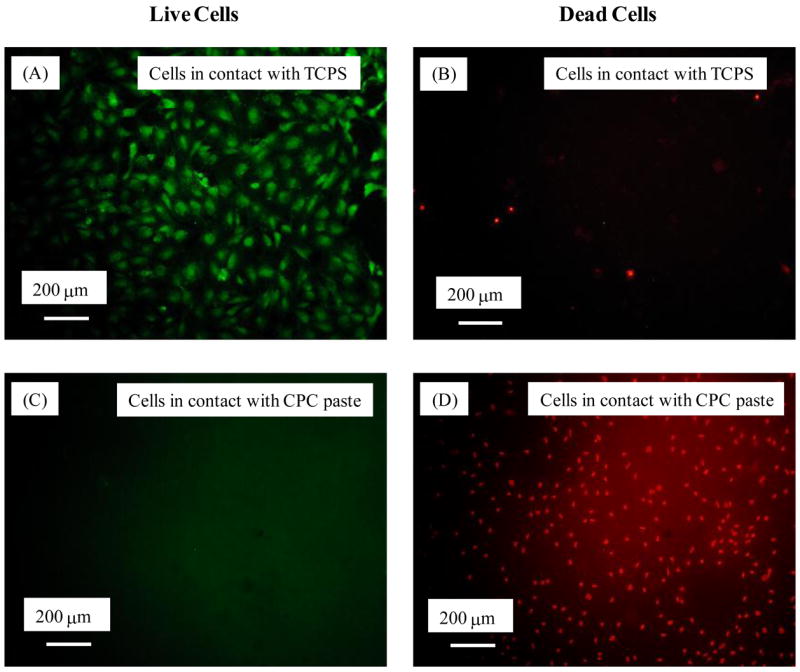
Methods: Tetracalcium phosphate [TTCP: Ca(4)(PO(4))(2)O] and dicalcium phosphate (DCPA: CaHPO(4)) were used to fabricate self-setting calcium phosphate cement. Strong and macroporous scaffolds were developed via absorbable fibers, biopolymer chitosan, and mannitol porogen. Following established protocols, MC3T3-E1 osteoblast-like cells (Riken, Hirosaka, Japan) were cultured on the specimens and inside the CPC composite paste carrier.

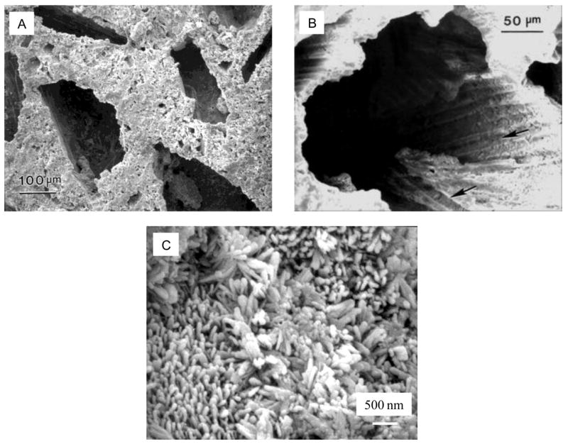
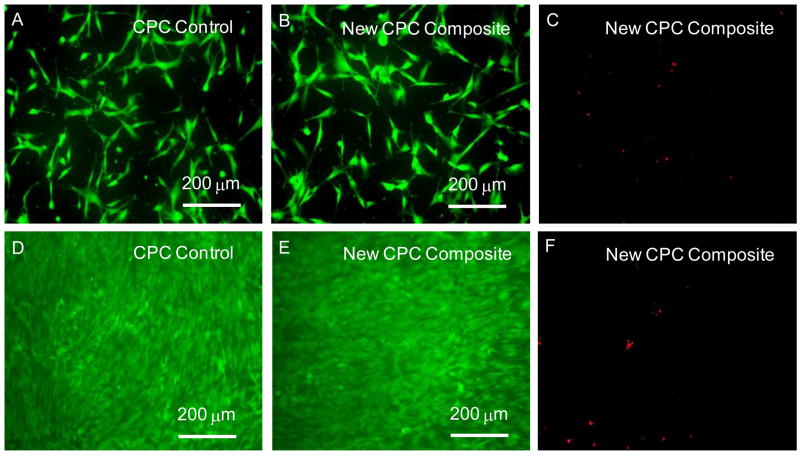
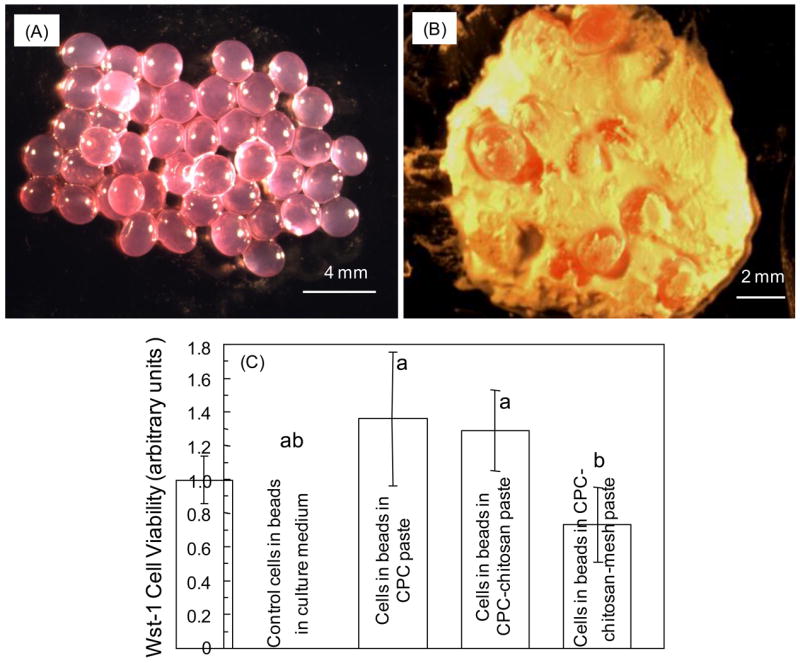
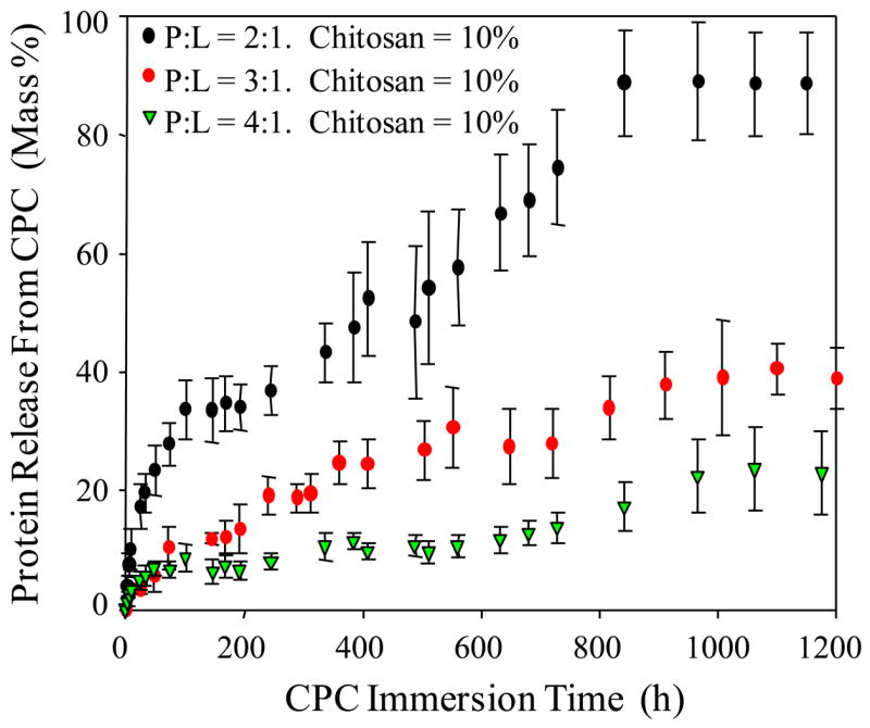
Results: The scaffold strength was more than doubled via reinforcement (p<0.05). Relationships and predictive models were established between matrix properties, fibers, porosity, and overall composite properties. The cement injectability was increased from about 60% to nearly 100%. Cell attachment and proliferation on the new composite matched those of biocompatible controls. Cells were able to infiltrate into the macropores and anchor to the bone mineral-like nano-apatite crystals. For cell delivery, alginate hydrogel beads protected cells during cement mixing and setting, yielding cell viability measured via the Wst-1 assay that matched the control without CPC (p>0.1). For growth factor delivery, CPC powder:liquid ratio and chitosan content provided the means to tailor the rate of protein release from CPC carrier.

Significance: New CPC scaffolds were developed that were strong, tough, macroporous and osteoconductive. They showed promise for injection in minimally invasive surgeries, and in delivering osteogenic cells and osteoinductive growth factors to promote bone regeneration. Potential applications include various dental, craniofacial, and orthopedic reconstructions.


Figures

Figure 1
Figure 1
(A) The first group of macropores was formed in CPC via the dissolution of mannitol during 1-day of immersion, while fibers in CPC provide the needed early strength. (B) The second group of macropores was formed in CPC by the dissolution of fibers after 17 weeks of immersion. The rational was that after being implanted for several weeks in vivo, new bone would have grown into the first group of macropores to increase the CPC strength. (C) Nano hydroxyapatite crystals that make up the CPC matrix.
Figure 2
Figure 2
Relationships between scaffold matrix property, fiber property, and composite property. (A) CPC composite-CPC matrix relationship. (B) CPC fiber composite-individual fiber relationship. (C) CPC scaffold strength-porosity volume fraction relationship. (D) Fracture toughness-porosity relationship. Each value is mean ± sd; n = 6. Each curve is the best fit to the experimental data as described in the text.
Figure 3
Figure 3
(A) The injectability for two different CPC pastes. Note the poor injectability without HPMC. Adding a small amount of HPMC dramatically improved the injectability. (B) The force required to inject the CPC paste containing mannitol porogen particles, which were later dissolved in a physiological solution to create macropores in the injectable CPC. The paste contained 1 % HPMC. The CPC was fully injectable having up to 40 % mannitol. The * in (B) indicates that the maximum force of 100 N was reached.
Figure 4
Figure 4
(A–C) After 1-day culture, live cells (stained green) appeared to have adhered and attained a normal polygonal morphology on the specimen. Dead cells (stained red) in (C) were very few on both materials. (D–F) After 14 days of culture, live cells had formed a confluent monolayer on all specimens. The cell density was much greater than the 1-d density, indicating that the cells had greatly proliferated. These results suggest that cell proliferation was similar, demonstrating that the new CPC composite was as non-cytotoxic as the FDA-approved CPC control and the TCPS control.
Figure 5
Figure 5
SEM micrographs showing cell infiltration into the macropores of CPC scaffolds. (A) The pore was large enough for the osteoblast cell “O”, and the cell had developed long cytoplasmic extensions “E” anchoring to the pore bottom. (B) Two cells had established cell-cell junction (arrow) in the pore. (C) High magnification shows secondary extensions sprouting from the primary extension “E”. (D) Cell extensions anchored to the nano-apatite crystals that make up the CPC matrix.
Figure 6
Figure 6
For cell delivery via the CPC paste carrier, the cement paste setting reaction was harmful to the cells, as shown in (C) and (D). However, once the cement was set, it was biocompatible and supported cell attachment and proliferation. Therefore, cell protection was needed during cement mixing and setting.
Figure 7
Figure 7
Cell delivery via CPC composite-hydrogel construct. (A) Cell-seeded alginate hydrogel beads. Cells were encapsulated into alginate beads which were then mixed into three pastes: conventional CPC, CPC-chitosan, and CPC-chitosan-mesh. (B) CPC paste mixed with the cell-seeded hydrogel beads. After 1-day culture inside the setting cements, there were numerous live cells and very few dead cells, indicating that the alginate beads adequately protected the cells. The cell viability (mean ± sd; n = 5) was measured using the Wst-1 assay. The absorbance at 450 nm was (1.36 ± 0.41) for the conventional CPC and (1.29 ± 0.24) for CPC-chitosan composite, similar to the (1.00 ± 0.14) for the control with the beads in the culture medium without any CPC (p > 0.1).
Figure 8
Figure 8
Protein release from CPC carrier for a growth factor release study. The cement powder:liquid ratio had a significant effect on protein release (p < 0.05). Powder:liquid ratio and chitosan content are shown to be key microstructural parameters that can be tailored to control the protein release profile from CPC to be application-specific [51].

References

    1. Praemer A, Furner S, Rice DP. Musculoskeletal conditions in the United States. Rosemont: Amer Acad Orthop Surg; 1999.
    1. Franceshi RT. Biological approaches to bone regeneration by gene therapy. Critical Reviews in Oral Bio Med. 2005;84:1093–1103. - PubMed
    1. Medical Data International. RP-651147 - Orthopedic and musculoskeletal markets: Biotechnology and tissue engineering. 2000. pp. 28–31.
    1. Ambrosio AMA, Sahota JS, Khan Y, Laurencin CT. A novel amorphous calcium phosphate polymer ceramic for bone repair: I. Synthesis and characterization. J Biomed Mater Res. 2001;58B:295–301. - PubMed
    1. Laurencin CT, Ambrosio AMA, Borden MD, Cooper JA. Tissue engineering: Orthopedic applications. Annual Rev Biomed Eng. 1999;1:19–46. - PubMed

Publication types

MeSH terms