Review
. 2008 Mar;28(2):120-32.
doi: 10.1016/j.semnephrol.2008.01.005.

New insights into the pathogenesis of idiopathic hypercalciuria

Affiliations
Review

New insights into the pathogenesis of idiopathic hypercalciuria

Elaine M Worcester et al. Semin Nephrol. 2008 Mar.

Abstract

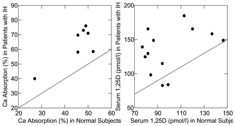
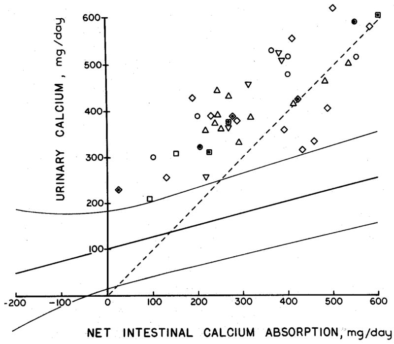
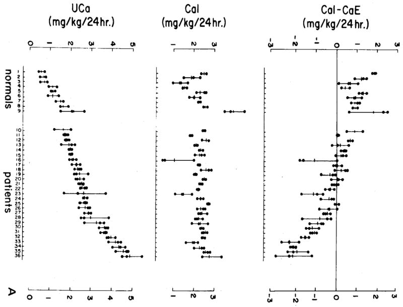
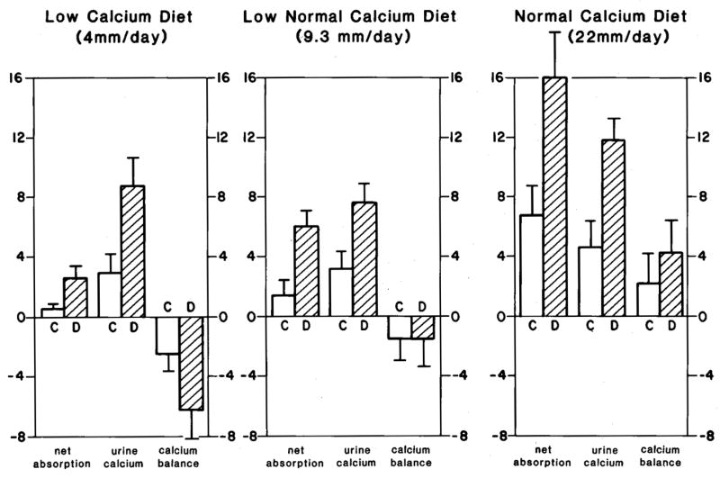
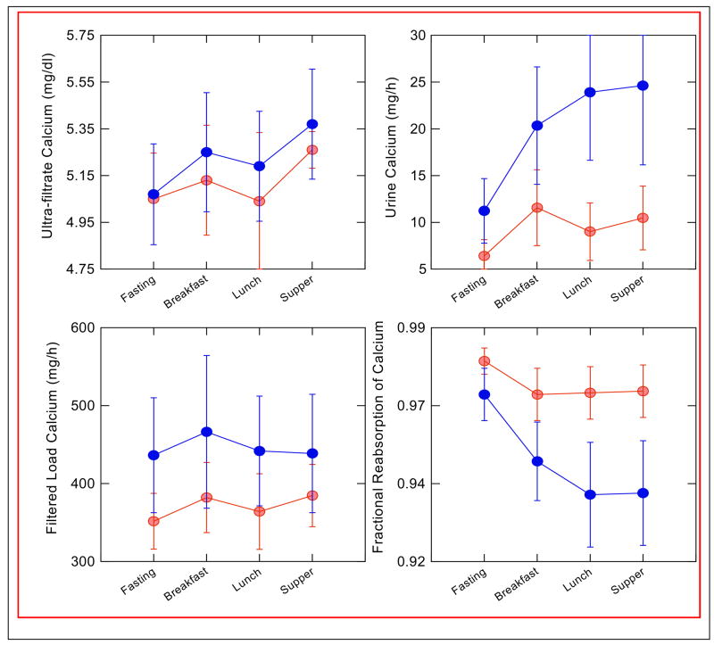
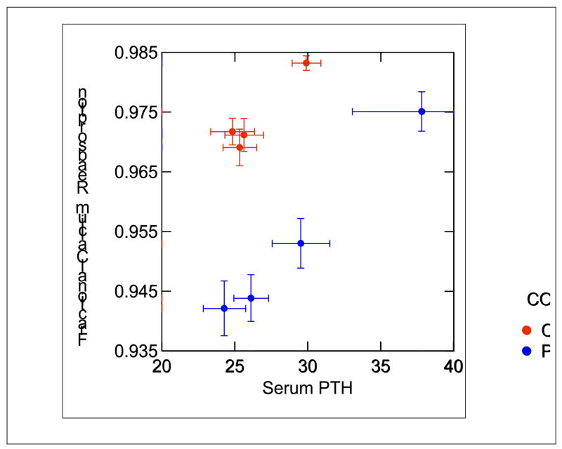
Idiopathic hypercalciuria (IH) is the most common metabolic abnormality in patients with calcium kidney stones. It is characterized by normocalcemia, absence of diseases that cause increased urine calcium, and calcium excretion that is greater than 250 mg/d in women and 300 mg/d in men. Subjects with IH have a generalized increase in calcium turnover, which includes increased gut calcium absorption, decreased renal calcium reabsorption, and a tendency to lose calcium from bone. Despite the increase in intestinal calcium absorption, a negative calcium balance is seen commonly in balance studies, especially on a low-calcium diet. The mediator of decreased renal calcium reabsorption is not clear; it is not associated with either an increase in filtered load of calcium or altered parathyroid hormone levels. There is an increased incidence of hypercalciuria in first-degree relatives of those with IH, but IH appears to be a complex polygenic trait with a large contribution from diet to expression of increased calcium excretion. Increased tissue vitamin D response may be responsible for the manifestations of IH in at least some patients.


Figures

Figure 1
Figure 1. Intestinal Calcium Absorption and Serum 1,25(OH)2D3 Levels in IH and Normals
Fractional absorption of radio-labeled calcium by patients with IH (y-axis, left panel) exceeds that of normal subjects (x-axis, left panel) in 7 studies as shown by the displacements of points about the diagonal line of identity. Each point compares mean values for IH vs. normal within a single study. The same was true for serum 1,25(OH)2D3 levels: those of IH patients (y-axis, right panel) exceeded normals (x-axis, right panel) in 11 of 13 studies. (From reference [10]).
Figure 2
Figure 2. Balance Studies of IH
In each of 42 metabolic balance studies of subjects with IH (individual laboratories reporting data are denoted by differing symbols) IH subjects excreted more calcium (y-axis) for any given level of net calcium absorption (x-axis) than did 203 normal subjects represented by the curving 95% confidence limits. Moreover, in all but 7 subjects, calcium excretion exceeded or equaled net calcium absorption as indicated by points lying above the diagonal dashed line of identity. (From reference [9]).
Figure 3
Figure 3. Response of IH and Normal Subjects to very Low Calcium Diet
Nine normal people and 27 people with IH ate a diet containing 2 mg/kg/day of calcium (middle panel) for 9 days; the actual amounts of calcium ingested during days 7–9 (middle panel) by normal and IH overlapped. Urine calcium excretion of IH subjects (lower panel) matched or exceeded the highest values for the normals, as did the difference between ingested and excreted calcium (upper panel, CaI – CaE). Many IH subjects excreted more calcium than they ingested, whereas no normal did so; the loss of calcium above diet content can only mean loss of bone mineral into urine. (From reference [16])
Figure 4
Figure 4. Response of Normal Men Given 1,25(OH)2D3 Supplements
On a low calcium diet (left panel) vitamin D (hatched bars) increased net calcium absorption and urine calcium excretion compared to control periods (clear bars), but because diet calcium was limiting urine calcium rose more than calcium absorption could and bone mineral balance became more negative than during the control periods. With 9.3 and 22 mM/day calcium diets (middle and right panels) calcium balance became neutral (middle panel) and positive (right panels) respectively. (From Coe FL, Parks JH: Nephrolithiasis: Pathogenesis and Treatment, 2nd ed. Chicago, Year Book Medical Publishers, 1988, p. 113. By permission.)
Figure 5
Figure 5. Response of IH Patients (Black circles) and Normals (Grey circles) to a Three Meal Day
Serum ultra-filtratable calcium rose with meals (upper left panel) but values did not differ between IH and normal, nor did filtered load of calcium (lower left panel). Urine calcium of IH rose far above normals, and fractional calcium reabsorption of IH was below normals. Therefore, response of IH to feeding was an abnormally marked fall in tubule calcium reabsorption that accounts for hypercalciuria. (From reference [28]).
Figure 6
Figure 6. Serum PTH in IH (Black circles) and Normal Subjects (Grey circles)
Fasting (upper right values for each group) and during meals, values of PTH did not differ in IH vs. normal. At all values of PTH, calcium reabsorption of IH was below normal (points are ± SEM). (From reference [28]).

References

    1. Hodkinson A, Pyrah LN. The urinary excretion of calcium and inorganic phosphate in 344 patients with calcium stone of renal origin. Br J Surg. 1958;48:10–18. - PubMed
    1. Coe FL. Treated and untreated recurrent calcium nephrolithiasis in patients with idiopathic hypercalciuria, hyperuricosuria, or no metabolic disorder. Ann Intern Med. 1977;87:404–410. - PubMed
    1. Flocks RH. Calcium and phosphorus excretion in the urine of patients with renal or ureteral calculi. JAMA. 1939;113:1466.
    1. Henneman PH, Benedict PH, Forbes AP, Dudley HR. Idiopathic Hypercalciuria. N Engl J Med. 1958;259:802–807. - PubMed
    1. van’t Hoff WG. Aetiological factors in paediatric urolithiasis. Nephron Clin Pract. 2004;98:45–48. - PubMed

Publication types