

The acute and long-term adverse effects of shock wave lithotripsy
- PMID: 18359401
- PMCID: PMC2900184
- DOI: 10.1016/j.semnephrol.2008.01.003
The acute and long-term adverse effects of shock wave lithotripsy
Abstract
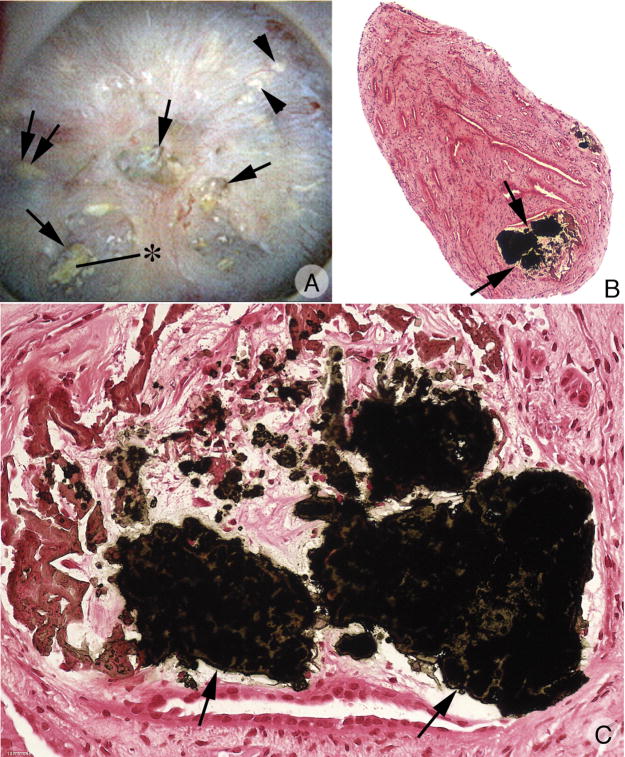
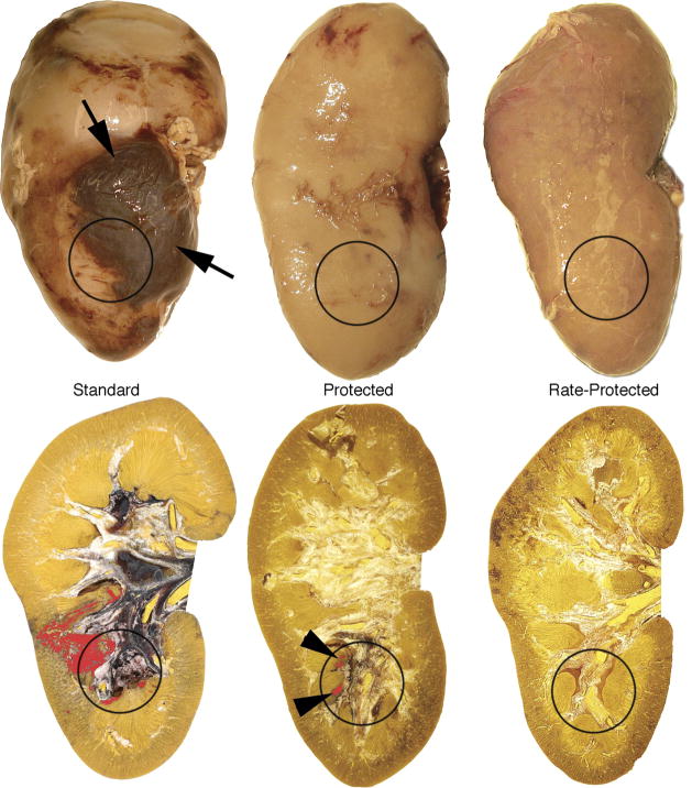
Shock wave lithotripsy (SWL) has proven to be a highly effective treatment for the removal of kidney stones. Shock waves (SWs) can be used to break most stone types, and because lithotripsy is the only noninvasive treatment for urinary stones, SWL is particularly attractive. On the downside SWL can cause vascular trauma to the kidney and surrounding organs. This acute SW damage can be severe, can lead to scarring with a permanent loss of functional renal volume, and has been linked to potentially serious long-term adverse effects. A recent retrospective study linking lithotripsy to the development of diabetes mellitus has further focused attention on the possibility that SWL may lead to life-altering chronic effects. Thus, it appears that what was once considered to be an entirely safe means to eliminate renal stones can elicit potentially severe unintended consequences. The purpose of this review is to put these findings in perspective. The goal is to explain the factors that influence the severity of SWL injury, update current understanding of the long-term consequences of SW damage, describe the physical mechanisms thought to cause SWL injury, and introduce treatment protocols to improve stone breakage and reduce tissue damage.
Figures
References
-
- Krambeck AE, Gettman MT, Rohlinger AL, Lohse CM, Patterson DE, Segura JW. Diabetes mellitus and hypertension associated with shock wave lithotripsy of renal and proximal ureteral stones at 19 years of followup. J Urol. 2006;175:1742–1747. - PubMed
-
- Lingeman JE, Matlaga B, Evan AP. Surgical management of urinary lithiasis. In: Walsh PC, Retik AB, Vaughan ED, Wein AJ, editors. Campbell’s Urology. Philadelphia: W.B Saunders Company; 2006. pp. 1431–1507. Chapter 44.
-
- Williams JC, Jr, Saw KC, Paterson RF, Hatt EK, McAteer JA, Lingeman JE. Variability of renal stone fragility in shock wave lithotripsy. Urology. 2003;61:1092–1097. - PubMed
-
- Carr LK, Honey JD’A, Jewett MAS, Ibanez D, Ryan M, Bombardier C. New stone formation: A comparision of extracorporeal shock wave lithotripsy and percutaneous nephrolithotomy. J Urol. 1996;155:1565–1567. - PubMed
-
- Fine JK, Pak CYC, Preminger GM. Effect of medical management and residual fragments on recurrent stone formation following shock wave lithotripsy. J Urol. 1995;153:27–33. - PubMed
Publication types
MeSH terms
Grants and funding
LinkOut - more resources
Full Text Sources
Other Literature Sources
