. 2008 Jul;95(1):378-89.
doi: 10.1529/biophysj.107.125229. Epub 2008 Mar 21.

Quantitative lifetime unmixing of multiexponentially decaying fluorophores using single-frequency fluorescence lifetime imaging microscopy

Affiliations

Quantitative lifetime unmixing of multiexponentially decaying fluorophores using single-frequency fluorescence lifetime imaging microscopy

Gert-Jan Kremers et al. Biophys J. 2008 Jul.

Abstract

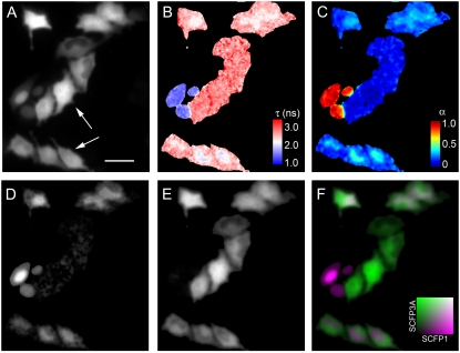
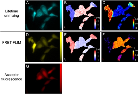
Fluorescence lifetime imaging microscopy (FLIM) is a quantitative microscopy technique for imaging nanosecond decay times of fluorophores. In the case of frequency-domain FLIM, several methods have been described to resolve the relative abundance of two fluorescent species with different fluorescence decay times. Thus far, single-frequency FLIM methods generally have been limited to quantifying two species with monoexponential decay. However, multiexponential decays are the norm rather than the exception, especially for fluorescent proteins and biological samples. Here, we describe a novel method for determining the fractional contribution in each pixel of an image of a sample containing two (multiexponentially) decaying species using single-frequency FLIM. We demonstrate that this technique allows the unmixing of binary mixtures of two spectrally identical cyan or green fluorescent proteins, each with multiexponential decay. Furthermore, because of their spectral identity, quantitative images of the relative molecular abundance of these fluorescent proteins can be generated that are independent of the microscope light path. The method is rigorously tested using samples of known composition and applied to live cell microscopy using cells expressing multiple (multiexponentially decaying) fluorescent proteins.


Figures

FIGURE 1
FIGURE 1
Excitation (dotted lines) and emission spectra (solid lines) of fluorescent proteins. (A) Spectra of SCFP1 (gray) and SCFP3A (black). (B) Spectra of SGFP2(T65G) (gray) and SGFP2 (black).
FIGURE 2
FIGURE 2
Lifetime unmixing of mixtures of purified fluorescent proteins. (A–C) Mixtures containing SCFP1 and SCFP3A. (D–G) Mixtures containing SGFP2(T65G) and SGFP2. α is the fractional contribution of SCFP1 or SGFP2(T65G) in each sample. (A and D) Comparison of the measured τϕ (○) and τm (▵) versus α. The solid lines represent the theoretical relation of τϕ (black) and τm (gray) with α. (B and E) Correlation between α (τϕ) and the true sample composition α. Solid lines are the linear fits, and dotted lines represent α (τϕ) = α. (C and F) Correlation between α (τm) versus the actual sample composition α. Solid lines are the linear fits, and dotted lines represent α (τm) = α. (G) Estimation of composition of SGFP2(T65G)/SGFP2 mixtures by global analysis. Dotted line represents α (global analysis) = α. The lifetime values used for unmixing were taken from Table 1. Error bars indicate standard deviation (n = 6).
FIGURE 3
FIGURE 3
Lifetime unmixing of SCFP1 and SCFP3A in HeLa cells. (A) Fluorescence micrograph of cells expressing SCFP1-NLS and SCFP3A-NES or SCFP1-NLS alone. Arrows indicate cells that cannot be distinguished, based on the distribution of fluorescence. (B) Lifetime map based on τϕ. (C) Map of the fractional contribution of SCFP1 to the steady-state fluorescence. (D and E) Micrographs of the fractional molar distribution of SCFP1-NLS and SCFP3A-NES, respectively. (F) Overlay of D and E, with SCFP1-NLS indicated in purple and SCFP3A-NES indicated in green. Lifetime values used for unmixing: SCFP3A τϕ = 2.47 ns, τm = 2.63 ns; SCFP1 τϕ = 1.23 ns, τm = 1.40 ns. Scale bar = 50 μm.
FIGURE 4
FIGURE 4
Translocation of a fluorescent biosensor for PtdIns(3,4,5)P3 synthesis, visualized by lifetime unmixing. Cells were imaged before (A–C) and after (D–F) 10 min stimulation with PDGF. (A and D) Fluorescence micrographs of cells expressing SGFP2(T65G) and SGFP2-NES-PHgrp1, respectively. (B and E) Lifetime maps based on phase lifetime (τϕ). (C and F) Maps of the fractional contribution of SGFP2-NES-PHgrp1 to the total fluorescence. Lifetime values used for unmixing: SGFP2 τϕ = 2.59 ns, τm = 2.66 ns; SGFP2(T65G) τϕ = 1.25 ns, τm = 1.30 ns. Scale bar = 50 μm.
FIGURE 5
FIGURE 5
Multiparameter FLIM, combining lifetime unmixing with FRET-FLIM. (A, D, and G) Fluorescence micrographs of steady-state fluorescence of SCFP1/SCFP3A, SYFP2, and mCherry fluorescence, respectively. (B) Lifetime image based on SCFP fluorescence. (C) Fractional distribution of SCFP1. (E) Lifetime image based on SYFP2 fluorescence. (F) FRET efficiency map based on the fluorescence lifetime of SYFP2. Lifetime values used for unmixing: SCFP3A τϕ = 2.47 ns, τm = 2.63 ns; SCFP1 τϕ = 1.23 ns, τm = 1.40 ns. Scale bar = 30 μm.

References

    1. Dumas, D., and J. F. Stoltz. 2005. New tool to monitor membrane potential by FRET voltage sensitive dye (FRET-VSD) using spectral and fluorescence lifetime imaging microscopy (FLIM). Interest in cell engineering. Clin. Hemorheol. Microcirc. 33:293–302. - PubMed
    1. Lin, H. J., P. Herman, and J. R. Lakowicz. 2003. Fluorescence lifetime-resolved pH imaging of living cells. Cytometry A. 52:77–89. - PMC - PubMed
    1. Gadella, T. W. Jr., T. M. Jovin, and R. M. Clegg. 1993. Fluorescence lifetime imaging microscopy (FLIM): spatial resolution of microstructures on the nanosecond time scale. Biophys. Chem. 48:221–239.
    1. Gadella, T. W. Jr., and T. M. Jovin. 1995. Oligomerization of epidermal growth factor receptors on A431 cells studied by time-resolved fluorescence imaging microscopy. A stereochemical model for tyrosine kinase receptor activation. J. Cell Biol. 129:1543–1558. - PMC - PubMed
    1. Wouters, F. S., P. I. Bastiaens, K. W. Wirtz, and T. M. Jovin. 1998. FRET microscopy demonstrates molecular association of non-specific lipid transfer protein (nsL-TP) with fatty acid oxidation enzymes in peroxisomes. EMBO J. 17:7179–7189. - PMC - PubMed

Publication types

MeSH terms

LinkOut - more resources