. 2008 Aug;95(4):1985-92.
doi: 10.1529/biophysj.107.128355. Epub 2008 Mar 21.

Lateral packing of mineral crystals in bone collagen fibrils

Affiliations

Lateral packing of mineral crystals in bone collagen fibrils

Christian Burger et al. Biophys J. 2008 Aug.

Abstract

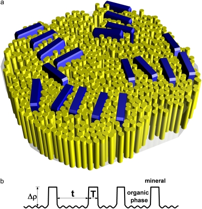
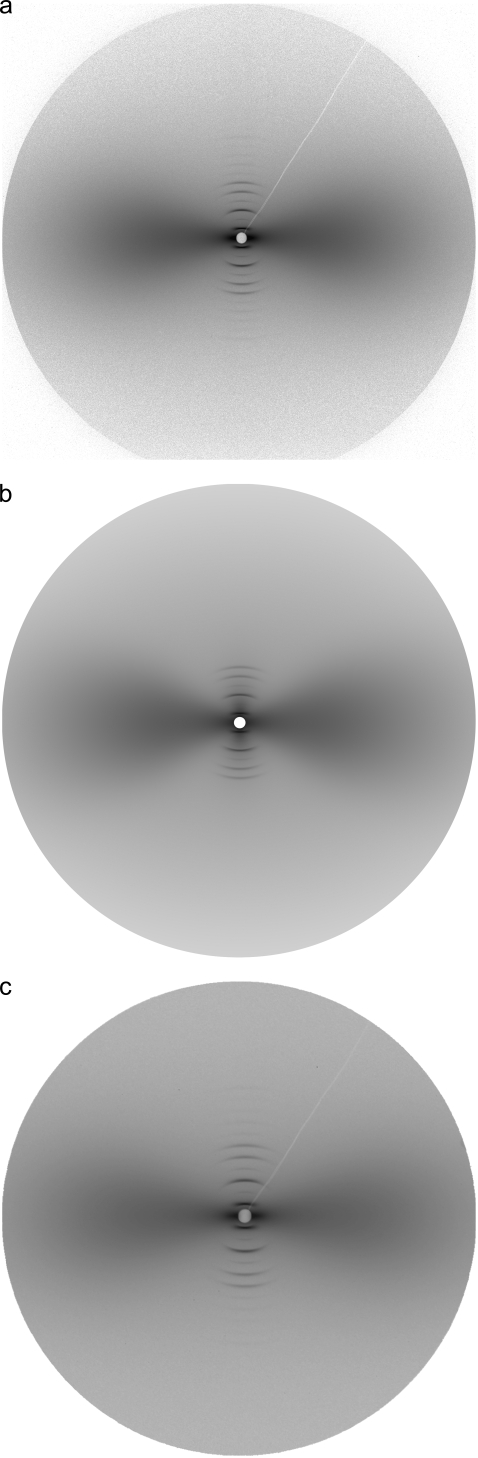
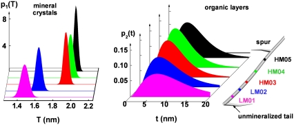
Combined small-angle x-ray scattering and transmission electron microscopy studies of intramuscular fish bone (shad and herring) indicate that the lateral packing of nanoscale calcium-phosphate crystals in collagen fibrils can be represented by irregular stacks of platelet-shaped crystals, intercalated with organic layers of collagen molecules. The scattering intensity distribution in this system can be described by a modified Zernike-Prins model, taking preferred orientation effects into account. Using the model, the diffuse fan-shaped small-angle x-ray scattering intensity profile, dominating the equatorial region of the scattering pattern, could be quantitatively analyzed as a function of the degree of mineralization. The mineral platelets were found to be very thin (1.5 nm approximately 2.0 nm), having a narrow thickness distribution. The thickness of the organic layers between adjacent mineral platelets within a stack is more broadly distributed with the average value varying from 6 nm to 10 nm, depending on the extent of mineralization. The two-dimensional analytical scheme also leads to quantitative information about the preferred orientation of mineral stacks and the average height of crystals along the crystallographic c axis.


Figures

FIGURE 1
FIGURE 1
(a) Schematic illustration of the lateral packing of mineral crystals in the collagen matrix. Thin apatite platelets are aligned nearly parallel within the stacks. The thickness of crystals is typically 2 nm and the width is ∼20 nm. The crystal height in the c-axis dimension (preferentially aligned about the fibril axis, not visible in this figure) is ∼30 nm. (b) Electron density projected onto the normal of a stack of mineral platelets. T is the thickness of individual crystals, and t is the thickness of organic layers between the neighboring crystals. The density of the mineral phase can be assumed to be uniform. There are small density fluctuations in the organic phase but their order of magnitude is much smaller than the density contrast (Δρ) between the mineral phase and the organic phase.
FIGURE 2
FIGURE 2
(a) Measured two-dimensional SAXS pattern from intramuscular shad bone by imaging plate. Sample/detector distance was 1897 mm and the measurement range covered s = 0.01 ∼ 0.38 nm−1. Bone axis is in the vertical direction (s3, meridian). The equator s12 is perpendicular to the bone axis. (b) Calculated pattern for panel a. The total intensity is a combination of the fan-shaped diffuse scattering originating from the lateral packing of mineral crystals in organic matrix (described in this article), and sharp meridional reflections originating from periodic packing of collagen molecules and mineral crystals along the fibril axis (see (28) for details). The corresponding fitting parameters are listed in Data S1, Table 1. (c) Diffraction pattern from the same irradiated volume as in panel a, but recorded with a CCD camera. Sample/detector distance was 1942 mm and the measurement range covered 0.01 ∼ 0.30 nm−1.
FIGURE 3
FIGURE 3
Evolution of the mineral crystal packing in the collagen matrix at different degrees of mineralization. The intramuscular shad bone sample (right) has roughly a fiber shape with an unmineralized tail at the end and a spur in the middle. The extent of mineralization gradually increases from the interface between the two portions (unmineralized and mineralized) to the spur. The thickness distributions of the mineral crystals are plotted as narrow Gaussians (Eq. 2) and the thickness distributions of the intercalated organic layers are plotted as Γ-distributions (Eq. 4). Note that the scale range for the mineral thickness distribution (left) is 1/20 of that for the organic layer thickness distribution (right).
FIGURE 4
FIGURE 4
TEM images of lightly mineralized intramuscular herring bone. Platelet-shaped mineral crystals are shown as dark lines in the cross section of the fibrils and form near parallel arrays schematically shown in Fig. 1. The scale bar is 100 nm.

References

    1. Glimcher, M. J. 1960. Specificity of the molecular structure of organic matrices in mineralization. In Calcification in Biological Systems. R. F. Sogannes, editor. American Association for the Advancement of Science, Washington, DC.
    1. Glimcher, M. J. 1984. Recent studies of the mineral phase in bone and its possible linkage to the organic matrix by protein-bound phosphate bonds. Philos. Trans. R. Soc. Lond. B Biol. Sci. 304:479–508. - PubMed
    1. Glimcher, M. J. 1998. The nature of the mineral phase in bone: Biological and clinical implications. In Metabolic Bone Disease and Clinically Related Disorders. L. V. Avioli, and S. M. Krane, editors. Academic Press, San Diego, CA.
    1. Glimcher, M. J. 2006. Bone: nature of the calcium phosphate crystals and cellular, structural, and physical chemical mechanisms in their formation. Rev. Mineral. Geochem. 64:223–282.
    1. Fitton-Jackson, S. 1957. The fine structure of developing bone in the embryonic fowl. Proc. Roy. Soc. (London) B. 14:270–280. - PubMed

Publication types