. 2008 Jul;95(2):641-7.
doi: 10.1529/biophysj.107.120808. Epub 2008 Mar 21.

Part II: diffraction from two-dimensional cholera toxin crystals bound to their receptors in a lipid monolayer

Affiliations

Part II: diffraction from two-dimensional cholera toxin crystals bound to their receptors in a lipid monolayer

C E Miller et al. Biophys J. 2008 Jul.

Abstract

The structure of cholera toxin (CTAB(5)) bound to its putative ganglioside receptor, galactosyl-N-acetylgalactosaminyl (N-acetyl-neuraminyl) galactosylglucosylceramide (GM(1)), in a lipid monolayer at the air-water interface has been studied utilizing grazing incidence x-ray diffraction. Cholera toxin is one of very few proteins to be crystallized in two dimensions and characterized in a fully hydrated state. The observed grazing incidence x-ray diffraction Bragg peaks indicated cholera toxin was ordered in a hexagonal lattice and the order extended 600-800 A. The pentameric binding portion of cholera toxin (CTB(5)) improved in-plane ordering over the full toxin (CTAB(5)) especially at low pH. Disulfide bond reduction (activation of the full toxin) also increased the protein layer ordering. These findings are consistent with A-subunit flexibility and motion, which cause packing inefficiencies and greater disorder of the protein layer. Corroborative out-of-plane diffraction (Bragg rod) analysis indicated that the scattering units in the cholera layer with CTAB(5) shortened after disulfide bond reduction of the A subunit. These studies, together with Part I results, revealed key changes in the structure of the cholera toxin-lipid system under different pH conditions.


Figures
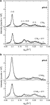
FIGURE 1
FIGURE 1
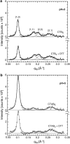
GIXD Bragg peaks obtained for layers of CTB5 and CTAB5 at pH = 8. The protein crystals nucleated under the DPPE:GM1 monolayer forming an ordered 2D protein monolayer with hexagonal packing. (a) Bragg peaks from CTB5 and CTB5 + DTT. Miller indices {h, k} of the observed peaks are indicated. Bragg peaks were integrated over the qz region (–0.05 to 0.2 Å−1). Peaks were fitted (solid lines) using Lorenzian curves (see Table 1 for details). (b) Bragg peaks from CTAB5 and CTAB5 + DTT. Intensities and positions of the Bragg peaks (especially higher order) in the case of CTAB5 had larger uncertainties due to weaker in-plane ordering and a higher incoherent background contribution due to the A subunit.
FIGURE 2
FIGURE 2
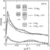
(a) Bragg rod profiles corresponding to scattering from the cholera protein layer at pH = 8. Rods were produced by integrating through the 0.08 Å−1qxy ≤ 0.12 Å−1 region of the {1,0} Bragg peaks in Fig. 1. The profiles corresponding to CTAB5 have been offset by 6 for clarity. The sharp peak at qxy∼0.01 Å−1 is the Vineyard-Yoneda peak (18). Error bars are smaller than the symbol size. Lines are fits to the profiles based on cylinders shown in b. (b) Schematic representation of the cylindrical objects used to approximate the scattering units. Structural parameters such as the cylinder's height, radius, and tilt angle of the cholera molecules are reported in Table 2. In the case of CTAB5, two populations of cylinders were used to fit the data. After activation of CTAB5 there was a shift from ∼75% to ∼40% for population 2.
FIGURE 3
FIGURE 3
Grazing incidence x-ray diffraction (GIXD) Bragg peaks obtained for layers of CTB5 and CTAB5 with a subphase at pH = 5. The crystals nucleated on the DPPE:GM1 monolayer forming an ordered 2D protein monolayer. The observed GIXD Bragg peaks indicate packing of the toxins in a hexagonal 2D unit cell. (a) Bragg peaks from CTB5 and CTB5 + DTT. Miller indices {h, k} of the observed peaks are indicated. Bragg peaks were integrated over the qz region from −0.05 to 0.2 Å−1. Peaks were fitted (solid lines) using Lorenzian curves (see Table 1 for details). (b) Bragg peaks from CTAB5 and CTAB5 + DTT. Intensities and positions of the Bragg peaks (especially higher order) in the case of CTAB5 had larger uncertainties due to weaker in-plane ordering and higher incoherent background contribution.
FIGURE 4
FIGURE 4
Bragg rod profiles corresponding to scattering from the cholera protein layer at pH = 5. Rods were produced by integrating through the 0.08 Å−1qxy ≤ 0.12 Å−1 region of the {1,0} Bragg peaks in Fig. 3. Cylindrical objects were used to approximate the scattering units. Structural parameters such as the cylinder's height, radius, and tilt angle of the cholera molecules are reported in Table 2. In the case of CTAB5, two populations of cylinders were used to fit the data. The curves corresponding to CTAB5 have been offset for clarity. The sharp peak at qxy = 0.01Å−1 is the Vineyard-Yoneda peak (18).

References

    1. Haas, H., G. Brezesinski, and H. Mohwald. 1995. X-ray-diffraction of a protein crystal anchored at the air-water-interface. Biophys. J. 68:312–314. - PMC - PubMed
    1. Lenne, P. F., B. Berge, A. Renault, C. Zakri, C. Venien-Bryan, S. Courty, F. Balavoine, W. Bergsma-Schutter, A. Brisson, G. Grubel, N. Boudet, O. Konovalov, and J. F. Legrand. 2000. Synchrotron radiation diffraction from two-dimensional protein crystals at the air/water interface. Biophys. J. 79:496–500. - PMC - PubMed
    1. Verclas, S. A. W., P. A. Howes, K. Kjaer, A. Wurlitzer, M. Weygand, G. Buldt, N. A. Dencher, and M. Losche. 1999. X-ray diffraction from a single layer of purple membrane at the air/water interface. J. Mol. Biol. 287:837–843. - PubMed
    1. Verclas, S. A. W., P. A. Howes, K. Kjaer, A. Wurlitzer, M. Weygand, G. Buldt, N. A. Dencher, and M. Losche. 1999. X-ray diffraction from a single layer of purple membrane at the air/water interface. J. Mol. Biol. 287:837–843. - PubMed
    1. Weygand, M., K. Kjaer, P. B. Howes, B. Wetzer, D. Pum, U. B. Sleytr, and M. Losche. 2002. Structural reorganization of phospholipid headgroups upon recrystallization of an S-layer lattice. J. Phys. Chem. B. 106:5793–5799.

Publication types

LinkOut - more resources