. 2008 Apr;17(4):725-35.
doi: 10.1110/ps.073355508.

Structure-function studies on the active site of the coelenterazine-dependent luciferase from Renilla

Affiliations

Structure-function studies on the active site of the coelenterazine-dependent luciferase from Renilla

Jongchan Woo et al. Protein Sci. 2008 Apr.

Abstract

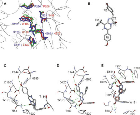
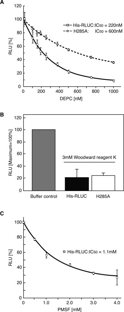
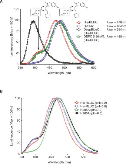
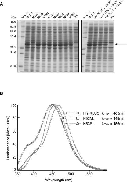
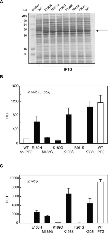
Renilla luciferase (RLUC) is a versatile tool for gene expression assays and in vivo biosensor applications, but its catalytic mechanism remains to be elucidated. RLUC is evolutionarily related to the alpha/beta hydrolase family. Its closest known homologs are bacterial dehalogenases, raising the question of how a protein with a hydrolase fold can function as a decarboxylating oxygenase. Molecular docking simulations with the coelenterazine substrate against an RLUC homology model as well as a recently determined RLUC crystal structure were used to build hypotheses to identify functionally important residues, which were subsequently tested by site-directed mutagenesis, heterologous expression, and bioluminescence emission spectroscopy. The data highlighted two triads of residues that are critical for catalysis. The putative catalytic triad residues D120, E144, and H285 bear only limited resemblance to those found in the active site of aequorin, a coelenterazine-utilizing photoprotein, suggesting that the reaction scheme employed by RLUC differs substantially from the one established for aequorin. The role of H285 in catalysis was further supported by inhibition using diethylpyrocarbonate. Multiple substitutions of N53, W121, and P220--three other residues implicated in product binding in the homologous dehalogenase Sphingomonas LinB--also supported their involvement in catalysis. Together with luminescence spectra, our data lead us to propose that the conserved catalytic triad of RLUC is directly involved in the decarboxylation reaction of coelenterazine to produce bioluminescence, while the other active-site residues are used for binding of the substrate.


Figures

Figure 1.
Figure 1.
RLUC protein structure displays and substrate docking simulations. (A) Overlay comparison between the catalytic triad and other active-site residues of Sphingomonas haloalkane dehalogenase, LinB (PDB IZ7A, red), the corresponding residues in the wild-type RLUC homology model (green), and the RLUC8 crystal structure (PDB 2PSD, blue) (Loening et al. 2007a). (B) Coelenterazine; oxygen and nitrogen are colored red and blue, respectively. Carbon and selected hydrogen are gray and white. (C–E) Docking simulations of coelenterazine to the lower portion of the RLUC active site including the putative catalytic triad. Hydrogen bonds are symbolized by green lines. (C,D) Docking of native coelenterazine (C) and the reaction intermediate, 2-hydroperoxy-coelenterazine (D) against the RLUC homology model. Note interactions between the hydroperoxy group and active-site residues N53, W121, and P220. (E) Docking of the reaction intermediate, 2-hydroperoxy-coelenterazine, was performed with the RLUC crystal structure obtained after exposure to substrate (PDB 2PSJ). In this alternative docking simulation, the reaction intermediate is suspended by hydrogen bonds between the R1 and R3 hydroxyls to N53 and the backbone of F262, respectively, while the reactive center is juxtaposed to the catalytic triad.
Figure 2.
Figure 2.
Inhibitor studies of RLUC. (A) DEPC. (B) Woodward Reagent K. (C) PMSF. Error bars represent standard error from n = 3 repeats. Assays were performed with 10 nM RLUC, preincubated with inhibitor at the indicated concentration for 30 min. The substrate concentration was 2 μM. In A, the activity of each protein in the absence of inhibitor was normalized to the peak value for ease of comparison. The H285A mutant has 11% of wild-type activity (Table 1).
Figure 3.
Figure 3.
Luminescence spectra. (A) Wild-type His-RLUC (10 nM, 2 μM native coelenterazine or 40 nM with 4 μM DeepBlueC) was compared with the H285A mutant (100 nM, 3.1 μM native coelenterazine). Samples were scanned in triplicate from 350 nm to 600 nm at 1 nm/sec and normalized to peak at 100%. Note that the spectra are distorted because of the loss of enzyme activity over the time of the scan (∼5 min), a necessary condition for highlighting the emission spectrum around 400 nm. The presumptive structures underlying emission at 400 nm (neutral coelenteramide) and 470 nm (phenolate anion) are shown. Control experiments were performed with wild-type His-RLUC by initiating the scan at different wavelengths to confirm that the shoulder at 400 nm is not specific to the early phase of the luminescence reaction (supplemental Fig. S3). (B) Wild-type RLUC (His-RLUC) and the H285A mutant RLUC with 2 μM native coelenterazine were scanned at pH 7.2 and pH 6.0.
Figure 4.
Figure 4.
Expression levels and luminescence spectra of representative mutant RLUC proteins. (A) Coomassie blue-stained polyacrylamide gel demonstrating equal accumulation of wild-type RLUC and several representative RLUC mutant proteins after 1 h of induction of expression with IPTG. The N53C mutant is a rare exception. (Right) A dilution series of RLUC extract in empty vector extract (EV). The arrow points to recombinant RLUC. (B) Luminescence spectra of two mutations affecting residue N53. The conditions are as for Figure 3.
Figure 5.
Figure 5.
Enzyme activities (relative light units) for wild-type RLUC and selected mutants. (A) Polyacrylamide gel for the RLUC expression strains shown in B. (B) Values are in vivo luminescence activities from E. coli strain BL21 after induction of RLUC with IPTG. (C) Values are in vitro luminescence activities of purified proteins in the presence of coelenterazine substrate. Note the similarity between the activity profiles (B) in vivo and (C) in vitro.

References

    1. Coulon, V., Audet, M., Homburger, V., Bockaert, J., Fagni, L., Bouvier, M., Perroy, J. Subcellular imaging of dynamic protein interactions by bioluminescence resonance energy transfer. Biophys. J. 2008;94:1001–1009. - PMC - PubMed
    1. Deng, L., Markova, S.V., Vysotski, E.S., Liu, Z.J., Lee, J., Rose, J., Wang, B.C. Crystal structure of a Ca2+-discharged photoprotein: Implications for mechanisms of the calcium trigger and bioluminescence. J. Biol. Chem. 2004;279:33647–33652. - PubMed
    1. Frerichs-Deeken, U., Ranguelova, K., Kappl, R., Huttermann, J., Fetzner, S. Dioxygenases without requirement for cofactors and their chemical model reaction: Compulsory order ternary complex mechanism of 1H-3-hydroxy-4-oxoquinaldine 2,4-dioxygenase involving general base catalysis by histidine 251 and single-electron oxidation of the substrate dianion. Biochemistry. 2004;43:14485–14499. - PubMed
    1. Hart, R.C., Matthews, J.C., Hori, K., Cormier, M.J. Renilla reniformis bioluminescence: Luciferase-catalyzed production of non-radiating excited states from luciferin analogues and elucidation of the excited state species involved in energy transfer to Renilla green fluorescent protein. Biochemistry. 1979;18:2204–2210. - PubMed
    1. Head, J.F., Inouye, S., Teranishi, K., Shimomura, O. The crystal structure of the photoprotein aequorin at 2.3 Å resolution. Nature. 2000;405:372–376. - PubMed

Publication types

MeSH terms

LinkOut - more resources