. 2008 Jun;294(6):L1049-54.
doi: 10.1152/ajplung.00526.2007. Epub 2008 Mar 21.

Toll-like receptor 2 is upregulated by hog confinement dust in an IL-6-dependent manner in the airway epithelium

Affiliations

Toll-like receptor 2 is upregulated by hog confinement dust in an IL-6-dependent manner in the airway epithelium

K L Bailey et al. Am J Physiol Lung Cell Mol Physiol. 2008 Jun.

Abstract

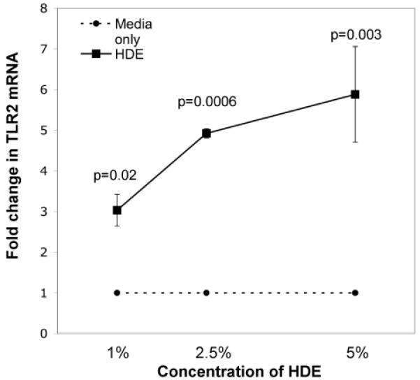
Hog confinement workers are at high risk to develop chronic bronchitis as a result of their exposure to organic dust. Chronic bronchitis is characterized by inflammatory changes of the airway epithelium. A key mediator in inflammation is Toll-like receptor 2 (TLR2). We investigated the role of TLR2 in pulmonary inflammation induced by hog confinement dust. Normal human bronchial epithelial cells (NHBE) were grown in culture and exposed to hog confinement dust extract. Hog confinement dust upregulated airway epithelial cell TLR2 mRNA in a concentration- and time-dependent manner using real-time PCR. There was a similar increase in TLR2 protein at 48 h as shown by Western blot. TLR2 was upregulated on the surface of airway epithelial cells as shown by flow cytometry. A similar upregulation of pulmonary TLR2 mRNA and protein was shown in a murine model of hog confinement dust exposure. Hog confinement dust is known to stimulate epithelial cells to produce IL-6. To determine whether TLR2 expression was being regulated by IL-6, the production of IL-6 was blocked using an IL-6-neutralizing antibody. This resulted in attenuation of the dust-induced upregulation of TLR2. To further demonstrate the importance of IL-6 in the regulation of TLR2, NHBE were directly stimulated with recombinant human IL-6. IL-6 alone was able to upregulate TLR2 in airway epithelial cells. Hog confinement dust upregulates TLR2 in the airway epithelium through an IL-6-dependent mechanism.


Figures

Figure 1
Figure 1
TLR2 mRNA is upregulated by hog confinement dust in a time- and concentration-dependent manner in airway epithelial cells. A. NHBE were incubated with (solid line) or without (dashed line) 5% HDE at 1,6,and 24 hours. Real-time PCR for TLR2 was performed. Data is expressed as fold change in TLR2 compared to control ± SEM. (n=4). B. NHBE were incubated with varying concentrations of HDE (solid line) or without (dashed line) for 24 hours. Real-time PCR for TLR2 was performed (n=4).
Figure 1
Figure 1
TLR2 mRNA is upregulated by hog confinement dust in a time- and concentration-dependent manner in airway epithelial cells. A. NHBE were incubated with (solid line) or without (dashed line) 5% HDE at 1,6,and 24 hours. Real-time PCR for TLR2 was performed. Data is expressed as fold change in TLR2 compared to control ± SEM. (n=4). B. NHBE were incubated with varying concentrations of HDE (solid line) or without (dashed line) for 24 hours. Real-time PCR for TLR2 was performed (n=4).
Figure 2
Figure 2
TLR2 protein is upregulated by hog confinement dust in the airway epithelium. A. NHBE were incubated with or without 5% HDE. Cells were lysed and Western blot for TLR2 is shown. TLR2 was detected at its expected molecular mass of, 86 kD. This is a representative blot of 4 separate experiments. B. Densitometry was performed showing a greater than 4 fold increase in TLR2 protein expressed in the HDE-treated cells compared to the media-treated cells. C. In the same experiments we also probed for TLR4. D. Densitometry showed no increase in TLR4 in response to HDE.
Figure 3
Figure 3
TLR2 receptors are upregulated on the plasma membrane by hog confinement dust in the airway epithelium. NHBE were incubated with media only, 1% HDE and 5% HDE for 48 hours. FACS staining for TLR2 was performed. A. Representative histogram of 3 separate experiments. Open histogram represents isotype control and solid histogram represents TLR2 staining. B. Summary bar graph shows an increasing Mean Fluorescence Intensity (MFI) with increasing concentrations of HDE.
Figure 4
Figure 4
TLR2 mRNA and protein is upregulated in the lung of a murine model of hog confinement dust exposure. A. Mice (n=4/group) were exposed to PBS, 1% HDE, 5% HDE and 12.5% HDE daily for 1 week. RNA was extracted from total lung homogenate and real-time PCR for TLR2 was performed. Data is expressed as mean fold change from mice with no handling ± SEM. B. Western blot for TLR2 from a representative mouse lung from each group demonstrates the 86kDa TLR2 protein band is upregulated in response to increasing concentrations of instilled HDE. C. Beta Actin loading control lane. D. Ratio of the densitometry of the TLR2 Western blot to the Beta Actin loading control demonstrates a 4-5 fold increase in TLR2 protein in response to mouse lung instillation with 1-12.5% HDE.
Figure 4
Figure 4
TLR2 mRNA and protein is upregulated in the lung of a murine model of hog confinement dust exposure. A. Mice (n=4/group) were exposed to PBS, 1% HDE, 5% HDE and 12.5% HDE daily for 1 week. RNA was extracted from total lung homogenate and real-time PCR for TLR2 was performed. Data is expressed as mean fold change from mice with no handling ± SEM. B. Western blot for TLR2 from a representative mouse lung from each group demonstrates the 86kDa TLR2 protein band is upregulated in response to increasing concentrations of instilled HDE. C. Beta Actin loading control lane. D. Ratio of the densitometry of the TLR2 Western blot to the Beta Actin loading control demonstrates a 4-5 fold increase in TLR2 protein in response to mouse lung instillation with 1-12.5% HDE.
Figure 5
Figure 5
IL-6 plays an important role in the upregulation of TLR2 in airway epithelial cells. A. NHBE were pre-incubated with or without anti-IL-6 for 1 hour then stimulated with or without 1% HDE for 24 hours. Real-time PCR for TLR2 was performed. Data expressed as fold change in TLR2 normalized to ribosomal RNA ± SEM. (n=4). B. NHBE were stimulated for 24 hours with varying concentrations of recombinant human IL-6. Real-time PCR for TLR2 was performed (n=4) showing that 20ng/ml of rhIL-6 significantly (p=0.01) increases TLR2 mRNA expression.
Figure 5
Figure 5
IL-6 plays an important role in the upregulation of TLR2 in airway epithelial cells. A. NHBE were pre-incubated with or without anti-IL-6 for 1 hour then stimulated with or without 1% HDE for 24 hours. Real-time PCR for TLR2 was performed. Data expressed as fold change in TLR2 normalized to ribosomal RNA ± SEM. (n=4). B. NHBE were stimulated for 24 hours with varying concentrations of recombinant human IL-6. Real-time PCR for TLR2 was performed (n=4) showing that 20ng/ml of rhIL-6 significantly (p=0.01) increases TLR2 mRNA expression.

References

    1. Basu S, Fenton MJ. Toll-like receptors: function and roles in lung disease. Am J Physiol Lung Cell Mol Physiol. 2004;286:L887–892. - PubMed
    1. Becker S, Dailey L, Soukup JM, Silbajoris R, Devlin RB. TLR-2 is involved in airway epithelial cell response to air pollution particles. Toxicol Appl Pharmacol. 2005;203:45–52. - PubMed
    1. Boasen J, Chisholm D, Lebet L, Akira S, Horner AA. House dust extracts elicit Toll-like receptor-dependent dendritic cell responses. J Allergy Clin Immunol. 2005;116:185–191. - PubMed
    1. Donham KJ, Scallon LJ, Popendorf W, Treuhaft MW, Roberts RC. Characterization of dusts collected from swine confinement buildings. Am Ind Hyg Assoc J. 1986;47:404–410. - PubMed
    1. Elliott MK, Sisson JH, West WW, Wyatt TA. Differential in vivo effects of whole cigarette smoke exposure versus cigarette smoke extract on mouse ciliated tracheal epithelium. Exp Lung Res. 2006;32:99–118. - PMC - PubMed

LinkOut - more resources