
The proteomes of human parotid and submandibular/sublingual gland salivas collected as the ductal secretions

Paul Denny et al. J Proteome Res. 2008 May.

Abstract

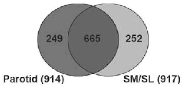
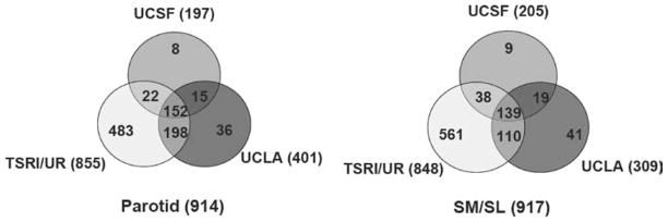
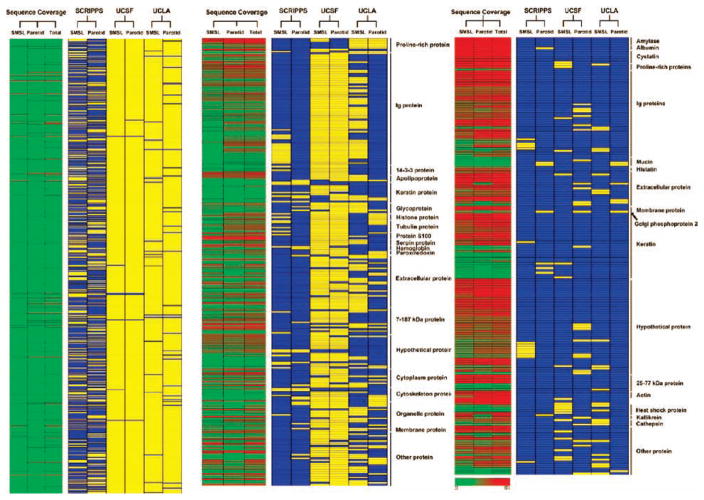
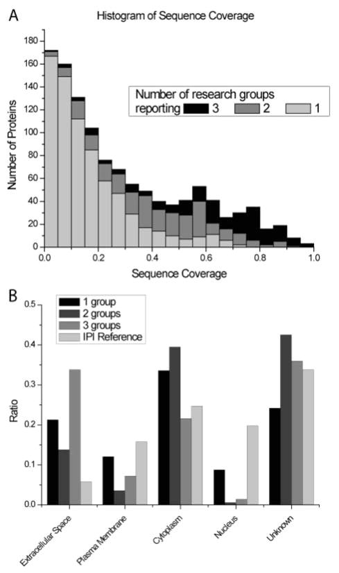
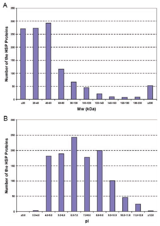
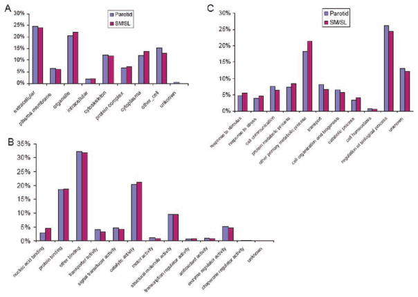
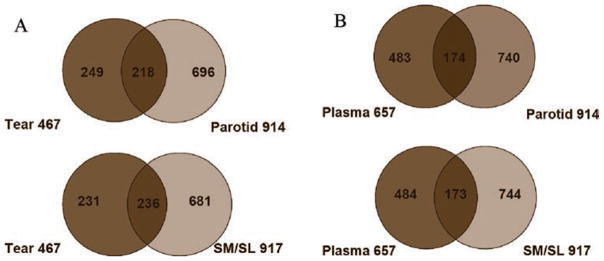
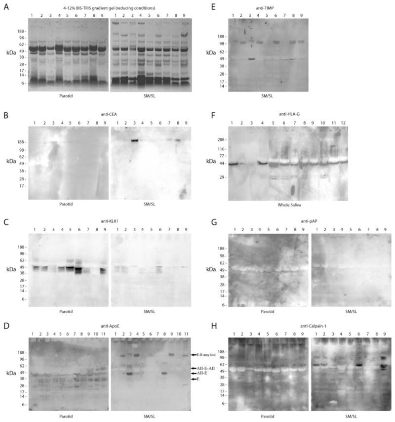
Saliva is a body fluid with important functions in oral and general health. A consortium of three research groups catalogued the proteins in human saliva collected as the ductal secretions: 1166 identifications--914 in parotid and 917 in submandibular/sublingual saliva--were made. The results showed that a high proportion of proteins that are found in plasma and/or tears are also present in saliva along with unique components. The proteins identified are involved in numerous molecular processes ranging from structural functions to enzymatic/catalytic activities. As expected, the majority mapped to the extracellular and secretory compartments. An immunoblot approach was used to validate the presence in saliva of a subset of the proteins identified by mass spectrometric approaches. These experiments focused on novel constituents and proteins for which the peptide evidence was relatively weak. Ultimately, information derived from the work reported here and related published studies can be used to translate blood-based clinical laboratory tests into a format that utilizes saliva. Additionally, a catalogue of the salivary proteome of healthy individuals allows future analyses of salivary samples from individuals with oral and systemic diseases, with the goal of identifying biomarkers with diagnostic and/or prognostic value for these conditions; another possibility is the discovery of therapeutic targets.


Figures

Figure 1
Figure 1
Human parotid and SM/SL salivas share many of the same protein components. The protein composition of each ductal fluid is presented as a Venn diagram. Fifty-seven percent of proteins were found in both parotid and submandibular/sublingual (SM/SL) salivas, whereas 27% were unique to the gland(s) of origin.
Figure 2
Figure 2
Venn diagrams illustrating the number of parotid (left) and SM/SL (right) proteins that were identified at each research site. Areas of overlap depict the number of proteins that were contributed by more than one group, with the core proteome—proteins that were identified by all three groups—shown in the center. Areas where the circles diverge show the number of protein identifications that were site-specific. Differences are likely attributable to the different methodologies employed by each research team.
Figure 3
Figure 3
Array view of the minimally overlapping human salivary proteome. Data were grouped according to the research team that identified each protein, the salivary gland where it was produced, and percent sequence coverage. The extent of coverage positively correlated with the number of research groups that identified a particular protein. The core proteome included one SM/SL-specific protein. Blue, protein identified; yellow, protein not identified; red, high-sequence coverage; yellow, intermediate-sequence coverage; and green, low-sequence coverage.
Figure 4
Figure 4
Sequence coverage and cellular origin of salivary proteins identified by single or multiple groups. (A) Histogram of the sequence coverage for proteins that were identified in parotid and SM/SL salivas. The shaded areas depict the number of proteins that were reported by one (gray), two (dark gray), or all three (black) research groups. (B) Comparison of the cellular location of salivary proteins identified by one, two, or all three research groups with a reference data set containing all human IPI database entries. Ratios of protein associations with the extracellular, plasma membrane, cytoplasm, or nuclear subcellular compartments were calculated using Ingenuity software. In some cases, this information was not known.
Figure 5
Figure 5
Molecular mass and pI distribution of the entire human salivary proteome. (A) The distribution was skewed to relatively low-molecular-mass components. (B) In contrast, the salivary proteins had a relatively broad range of pIs.
Figure 6
Figure 6
Relative allocation of the proteins identified in parotid and SM/SL saliva according to their gene ontology annotations. Components of both fluids were similarly distributed with regard to cellular locations (A), molecular functions (B), and biological processes (C). A high percentage of salivary proteins are predicted to be extracellular components or to reside within organelles (A). Salivary proteins were commonly involved in binding and catalysis (B). Parotid and SM/SL proteins had the highest distribution in metabolic and regulatory pathways (C).
Figure 7
Figure 7
Human parotid and SM/SL gland secretions share many protein components with human tears and plasma. (A) Areas of overlap depict the number of proteins that were identified in both tears and parotid (top) or SM/SL saliva (bottom). (B) Similarly, parotid (top) and SM/SL gland secretions (bottom) contained a substantial number of proteins that are also found in plasma.
Figure 8
Figure 8
Detection of novel salivary proteins and those with low sequence coverage. An immunoblot approach was used to confirm the presence in saliva of a portion of the proteins that were identified by using MS approaches. Most of the analyses shown employed parotid and SM/SL saliva samples collected as the ductal secretions from 5 females (lanes 1–5) and 4 males (lanes 6–9). As shown in panel F, whole saliva samples were collected from 10 females (lanes 1, 4–12) and 2 males (lanes 2–3). Proteins were selected for validation based on the number of research groups that reported the identification and the extent of sequence coverage. In general, emphasis was placed on the analysis of novel components and proteins with the least MS evidence of their presence in ductal saliva. (A) Samples were separated on 4–12% BIS-TRIS gradient gels under reducing conditions, and proteins were visualized by staining with Coomassie brilliant blue. Immunoblots detected the following: (B) CEA (IPI00027486, 6% sequence coverage, 1 group reporting), (C) kallikrein-1 (IPI00304808, 89% sequence coverage, 2 groups reporting), (D) apoE (IPI00021842, 21% sequence coverage, 1 group reporting), (E) TIMP-1 (SM/SL only, IPI00032292, 67% sequence coverage, 2 groups reporting), (F) HLA-G alpha chain, a nonclassical class I histocompatibility antigen (whole saliva, IPI00015988; a single peptide was sequenced by 1 research group), (G) prostatic acid phosphatase (IPI00396434, 8% sequence coverage, 1 group reporting), and (H) calpain-1 catalytic subunit (IPI00011285, 15% sequence coverage, 1 group reporting). E, apoE; AII-E, a complex of apoAII and apoE; AII-E-AII, AII-E with an additional apoAII; E-β-amyloid, a complex of apoE and β-amyloid.

References

    1. Levine MJ. Ann NY Acad Sci. 1993;694:11–16. - PubMed
    1. Lamkin MS, Oppenheim FG. Crit Rev Oral Biol Med. 1993;4:251–259. - PubMed
    1. Mandel ID. J Dent Res. 1987;66:623–627. - PubMed
    1. Humphrey SP, Williamson RT. J Prosthet Dent. 2001;85:162–169. - PubMed
    1. Dodds MW, Johnson DA, Yeh CK. J Dent. 2005;33:223–233. - PubMed

Publication types