Multivariate model of refractive shift in Descemet-stripping automated endothelial keratoplasty
- PMID: 18361978
- PMCID: PMC2796246
- DOI: 10.1016/j.jcrs.2007.11.045
Multivariate model of refractive shift in Descemet-stripping automated endothelial keratoplasty
Abstract
Purpose: To relate in situ graft shape in Descemet-stripping automated endothelial keratoplasty (DSAEK) to surgically induced refractive error.
Setting: Academic eye institute.
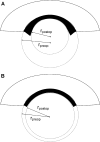
Methods: High frequency arc-scanning ultrasound was performed in 7 patients enrolled in a prospective study of microkeratome-assisted endothelial keratoplasty approved by the Investigative Review Board. A region of interest spanning the horizontal meridian was defined for analysis of epithelial, host, graft, and total corneal thicknesses. Graft thickness profiles were fit by quadratic polynomials where the 2nd-order coefficients represent the posterior corneal curvature contributed by the graft. The curvature coefficient and central graft thickness were analyzed as predictors of induced refractive error.
Results: At final follow-up (mean 5.9 months +/- 3.2 [SD]), 3 patients had a hyperopic shift (+2.50 diopters [D] each), 3 had insignificant (< 0.50 D) refractive shifts, and 1 had a myopic shift. In the group with hyperopic shift, a negative lens effect was predicted by positive curvature coefficients, representing grafts that were thinner centrally than peripherally (mean +22.72 microm/mm(2); range +4.95 to +45.17 microm/mm(2)). In the group with minimal refractive shift, coefficients were less positive (mean +7.28 microm/mm(2); range +2.01 to +13.82 microm/mm(2)). The patient with a myopic shift (-1.00 D) had the only negative curvature coefficient (-0.64 microm/mm(2)). In a 2-predictor model of refractive shift, central graft thickness and the curvature coefficient together accounted for 86% of the variance in the refractive response to DSAEK (P = .025).
Conclusion: Nonuniform thickness profiles and variable central graft thicknesses both contribute to refractive shift after DSAEK.
Conflict of interest statement
No author has a financial or proprietary interest in any material or method mentioned.
Figures


Similar articles
-
Effect of the shape of the endothelial graft on the refractive results after Descemet's stripping with automated endothelial keratoplasty.Can J Ophthalmol. 2009 Oct;44(5):557-61. doi: 10.3129/i09-087. Can J Ophthalmol. 2009. PMID: 19789591
-
Pentacam assessment of posterior lamellar grafts to explain hyperopization after Descemet's stripping automated endothelial keratoplasty.Ophthalmology. 2009 Sep;116(9):1651-5. doi: 10.1016/j.ophtha.2009.04.035. Epub 2009 Jul 29. Ophthalmology. 2009. PMID: 19643500
-
Visual acuity, refractive error, and endothelial cell density six months after Descemet stripping and automated endothelial keratoplasty (DSAEK).Cornea. 2007 Jul;26(6):670-4. doi: 10.1097/ICO.0b013e3180544902. Cornea. 2007. PMID: 17592314
-
[Corneal power after descemet stripping automated endothelial keratoplasty (DSAEK) - Modeling and concept for calculation of intraocular lenses].Z Med Phys. 2016 Jun;26(2):120-6. doi: 10.1016/j.zemedi.2015.02.003. Epub 2015 Mar 17. Z Med Phys. 2016. PMID: 25791739 Review. German.
-
Determinants of visual quality after endothelial keratoplasty.Surv Ophthalmol. 2016 May-Jun;61(3):257-71. doi: 10.1016/j.survophthal.2015.12.006. Epub 2015 Dec 19. Surv Ophthalmol. 2016. PMID: 26708363 Review.
Cited by
-
Accuracy of formulas for intraocular lens power for eyes undergoing descemet stripping automated endothelial keratoplasty and cataract surgery.Eye (Lond). 2024 Nov;38(16):3132-3135. doi: 10.1038/s41433-024-03242-7. Epub 2024 Jul 16. Eye (Lond). 2024. PMID: 39014210
-
Efficacy and safety of Descemet's membrane endothelial keratoplasty versus Descemet's stripping endothelial keratoplasty: A systematic review and meta-analysis.PLoS One. 2017 Dec 18;12(12):e0182275. doi: 10.1371/journal.pone.0182275. eCollection 2017. PLoS One. 2017. PMID: 29252983 Free PMC article.
-
Comparison of Artificial Anterior Chamber Internal Pressures and Cutting Systems for Descemet's Stripping Automated Endothelial Keratoplasty.Transl Vis Sci Technol. 2018 Nov 27;7(6):11. doi: 10.1167/tvst.7.6.11. eCollection 2018 Nov. Transl Vis Sci Technol. 2018. PMID: 30510855 Free PMC article.
-
Phacoemulsification in the Setting of Corneal Endotheliopathies: A Review.Int Ophthalmol Clin. 2020 Summer;60(3):71-89. doi: 10.1097/IIO.0000000000000315. Int Ophthalmol Clin. 2020. PMID: 32576725 Free PMC article. Review.
-
Sheets glide-assisted versus Busin glide-assisted insertion techniques for descemet stripping endothelial keratoplasty (DSEK): A comparative analysis.Med J Armed Forces India. 2019 Oct;75(4):370-374. doi: 10.1016/j.mjafi.2018.02.007. Epub 2018 Apr 5. Med J Armed Forces India. 2019. PMID: 31719729 Free PMC article.
References
-
- Gorovoy MS. Descemet-stripping automated endothelial keratoplasty. Cornea. 2006;25:886–889. - PubMed
-
- Price FW, Jr, Price MO. Descemet's stripping with endothelial keratoplasty in 50 eyes: a refractive neutral corneal transplant. J Refract Surg. 2005;21:339–345. - PubMed
-
- Price FW, Jr, Price MO. Descemet's stripping with endothelial keratoplasty in 200 eyes; early challenges and techniques to enhance donor adherence. J Cataract Refract Surg. 2006;32:411–418. - PubMed
-
- Koenig SB, Covert DJ, Dupps WJ, Jr, Meisler DM. Visual acuity, refractive error, and endothelial cell density six months after Descemet stripping and automated endothelial keratoplasty (DSAEK) Cornea. 2007;26:670–674. - PubMed
-
- Covert DJ, Koenig SB. New triple procedure: Descemet's stripping and automated endothelial keratoplasty combined with phacoemulsification and intraocular lens implantation. Ophthalmology. 2007;114:1272–1277. - PubMed

