. 2008 May 30;283(22):15390-8.
doi: 10.1074/jbc.M801589200. Epub 2008 Mar 24.

Mapping of POP1-binding site on pyrin domain of ASC

Affiliations

Mapping of POP1-binding site on pyrin domain of ASC

Thiagarajan Srimathi et al. J Biol Chem. .

Abstract

Apoptosis-associated speck-like protein containing a caspase recruitment domain (ASC) is an essential adaptor protein in the formation of a multiprotein complex that activates procaspase-1. ASC is also known as a modulator of NF-kappaB activation pathways. ASC has a bipartite domain structure, consisting of an N-terminal pyrin domain (PYD) and a C-terminal caspase-recruitment domain. The PYD of ASC (ASC_PYD) is known to interact with various PYD-containing intracellular danger signal sensors and PYD-only proteins. Using purified proteins, we characterized the in vitro interaction of ASC_PYD with PYD-only protein 1 (POP1). POP1 specifically interacts with ASC_PYD with a dissociation constant of 4.08 +/- 0.52 microm but does not interact with Cryopyrin. NMR and mutagenesis experiments show that a negative electrostatic potential surface patch (EPSP) on ASC_PYD, consisting of the first (H1) and fourth (H4) helices, is essential in the interaction with POP1. A positive EPSP on POP1, consisting of the second (H2) and third (H3) helices, is a counterpart of this interaction. The interaction between ASC_PYD and POP1 is similar to the interaction between caspase recruitment domains of Apaf-1 and procaspase-9. In addition, we present evidence that conformational changes at the long loop of ASC_PYD between the H2 and H3 helices can affect its interaction with POP1. Based on our observations, we propose that the positive EPSP of ASC_PYD, including the H2 and H3 helices, may be the binding site for Cryopyrin, and the interaction with Cryopyrin may induce the dissociation of POP1 from ASC_PYD.


Figures

FIGURE 1.
FIGURE 1.
Structure of ASC_PYD. Amino acid residues involved in the suggested negative (left) and positive (right) EPSPs are labeled. The electrostatic potentials mapped on solvent-accessible surface for each orientation are presented at the bottom. Positive and negative electrostatic potentials are shown in blue and red, respectively (±4 kT/e).
FIGURE 2.
FIGURE 2.
Amino acid sequence alignment of selected human and viral PYDs. Listed are the PYDs of human ASC (Swiss-Prot Q9ULZ3), human POP1 (Q8WXC3), human Cryo (Q96P20), human Pyrin (O15553), human POP2 (Q56P42), human NALP1 (Q9C000), human NALP2 (Q9NX02), human NALP5 (P59047), human NALP7 (Q8WX94), human AIM2 (O14862), rabbit fibroma virus GP013L_PYD (Q9Q957), myxoma virus M013L_PYD (Q9Q8S8), and swinepox virus SPV013 (Q8V3S3).
FIGURE 3.
FIGURE 3.
A, CD spectra of 10 μm purified ASC_PYD, POP, and Cryo_PYD are presented. B, size exclusion chromatography profiles of purified ASC_PYD (300 μm), POP1 (300 μm), and Cryo_PYD (60 μm) at pH 4.5.
FIGURE 4.
FIGURE 4.
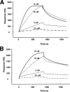
SPR binding assays. A, SPR sensograms to measure the KD for the interaction between ASC_PYD and POP1. ASC_PYD was immobilized on the sensor chip and various concentrations of POP1 (2.5-15 μm) were injected. B, SPR sensograms to measure the KD for the interaction between ASC_PYD_L25A and POP1.
FIGURE 5.
FIGURE 5.
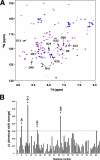
Chemical shift changes of ASC_PYD_L25A upon the addition of POP1. A, 1H-15N HSQC of 15N-labeled ASC_PYD alone (red) and with POP1 (blue) are overlaid. Resonance peaks of amino acid residues in the negative and positive EPSPs on ASC_PYD are labeled. B, a graph of the chemical shift change (Δδ) versus residue number.
FIGURE 6.
FIGURE 6.
Structure of POP1. Amino acid residues involved in the suggested negative (left) and positive (right) EPSPs are labeled. The electrostatic potentials mapped on the solvent-accessible surface for each orientation are presented at the bottom. Positive and negative electrostatic potentials are shown in blue and red, respectively (±4 kT/e).
FIGURE 7.
FIGURE 7.
Model structure of ASC_PYD·POP1 complex. Upper panel, ASC_PYD is shown in a surface presentation with electrostatic potential. POP1 is shown in a schematic diagram as salmon color. The amino acid residues of POP1 involved in the interaction are shown in a stick presentation. Bottom panel, POP1 is shown in a surface presentation with electrostatic potential. ASC_PYD is shown in a schematic diagram as green color.
FIGURE 8.
FIGURE 8.
Model structure of Cryo_PYD and electrostatic potential isosurfaces. The amino acid residues that affect the isosurfaces are labeled. Electrostatic potential isosurface (±2 kT/e) of Cryo_PYD is presented at the same orientation with the schematic diagram (top) and after 180° rotation (bottom). Positive and negative electrostatic potentials are shown in blue and red, respectively.

References

    1. Masumoto, J., Taniguchi, S., Ayukawa, K., Sarvotham, H., Kishino, T., Niikawa, N., Hidaka, E., Katsuyama, T., Higuchi, T., and Sagara, J. (1999) J. Biol. Chem. 274 33835-33838 - PubMed
    1. McConnell, B. B., and Vertino, P. M. (2004) Apoptosis 9 5-18 - PubMed
    1. Srinivasula, S. M., Poyet, J. L., Razmara, M., Datta, P., Zhang, Z., and Alnemri, E. S. (2002) J. Biol. Chem. 277 21119-21122 - PubMed
    1. Martinon, F., and Tschopp, J. (2007) Cell Death Differ. 14 10-22 - PubMed
    1. Agostini, L., Martinon, F., Burns, K., McDermott, M. F., Hawkins, P. N., and Tschopp, J. (2004) Immunity 20 319-325 - PubMed

Publication types

MeSH terms