. 2008 May 16;283(20):13688-99.
doi: 10.1074/jbc.M710499200. Epub 2008 Mar 24.

Regulatory and structural differences in the Cu,Zn-superoxide dismutases of Salmonella enterica and their significance for virulence

Affiliations

Regulatory and structural differences in the Cu,Zn-superoxide dismutases of Salmonella enterica and their significance for virulence

Serena Ammendola et al. J Biol Chem. .

Abstract

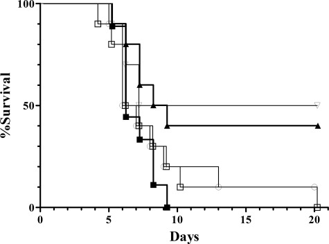
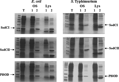
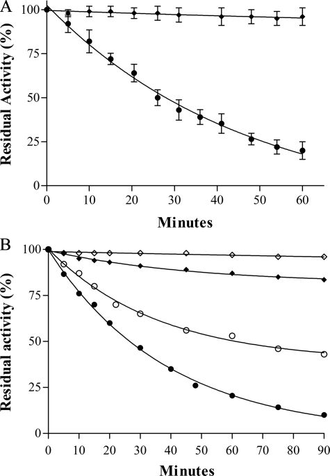
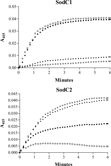
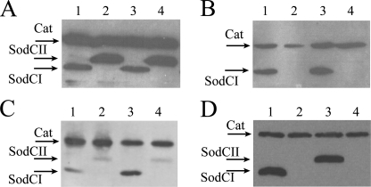
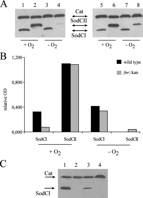
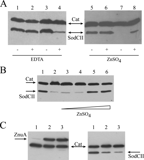
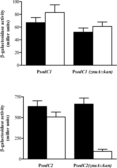
Many of the most virulent strains of Salmonella enterica produce two distinct Cu,Zn-superoxide dismutases (SodCI and SodCII). The bacteriophage-encoded SodCI enzyme makes the greater contribution to Salmonella virulence. We have performed a detailed comparison of the functional, structural, and regulatory properties of the Salmonella SodC enzymes. Here we demonstrate that SodCI and SodCII differ with regard to specific activity, protease resistance, metal affinity, and peroxidative activity, with dimeric SodCI exhibiting superior stability and activity. In particular, monomeric SodCII is unable to retain its catalytic copper ion in the absence of zinc. We have also found that SodCI and SodCII are differentially affected by oxygen, zinc availability, and the transcriptional regulator FNR. SodCII is strongly down-regulated under anaerobic conditions and dependent on the high affinity ZnuABC zinc transport system, whereas SodCI accumulation in vitro and within macrophages is FNR-dependent. We have confirmed earlier findings that SodCII accumulation in intracellular Salmonella is negligible, whereas SodCI is strongly up-regulated in macrophages. Our observations demonstrate that differences in expression, activity, and stability help to account for the unique contribution of the bacteriophage-encoded SodCI enzyme to Salmonella virulence.


Figures

FIGURE 1.
FIGURE 1.
Virulence of wild type and sodC mutant S. typhimurium in C3H/HeN mice. Survival is shown for groups of 10 mice infected with wild type (▪), sodCII::kan (○), sodCII::phoA (□), sodCII::pRR10(ΔtrfA)(▴), and sodCI::kan (▿) strains.
FIGURE 2.
FIGURE 2.
SodCI and SodCII release from the periplasm of E. coli and S. typhimurium. SDS-PAGE of total cellular lysates (T), pellet (lane 1) and periplasmic fractions (lane 2) released by osmotic shock (OS) or lysozyme treatment (Lys) from E. coli 71/18 or S. typhimurium ATTC14028 overexpressing SodCI, SodCII, or P. leiognathi SodC.
FIGURE 3.
FIGURE 3.
A, proteinase K susceptibility of SodCI and SodCII. Proteins were incubated at 37 °C in 20 mm Tris-HCl at pH 8.0, in the presence of 0.1 mg/ml proteinase K. Aliquots were withdrawn at the indicated times and assayed for residual SOD activity by the pyrogallol method. SodCI (♦), SodCII (•). B, EDTA inactivation of SodCI and SodCII. Cu,Zn-SOD samples at a concentration of 0.04 mg/ml were incubated at 37 °C in 100 mm phosphate buffer, 0.1 mm EDTA, pH 6.2 and 7.8. Aliquots were withdrawn at the indicated times and assayed for residual SOD activity as above. Each data point represents the mean of at least three independent measures. SodCI, pH 7.8 (♦), SodCII, pH 7.8 (•), SodCI, pH 6.2 (⋄), SodCII, pH 6.2 (○).
FIGURE 4.
FIGURE 4.
Peroxidase activity of SodCI and SodCII. The time-dependent increase in formula image formation was monitored at 415 nm as a measure of the peroxidase activity of SodCI and SodCII at 25 °C in the presence of 10 mm NaHCO3. Holo-enzyme (♦), holo-enzymes plus EDTA (⋄), zinc-free enzyme (•), and zinc-free enzyme plus EDTA (○) are shown. All bacterial enzymes showed no detectable peroxidative under the conditions tested in the absence of NaHCO3.
FIGURE 5.
FIGURE 5.
In vitro and in vivo SodCI and SodCII accumulation. Bacterial lysates were loaded on a 12% polyacrylamide gel, which was processed for Western blot analysis. Membranes were probed with anti-FLAG monoclonal antibodies. Strains MA7224 (lane 1), MA7225 (lane 2), SA167 (lanes 3), and SA168 (lane 4) were grown overnight in LB medium (panel A) or collected from J774.1 macrophages 23 h post-infection (panel B). Panel C, strains MA7224 (lanes 1 and 3) and MA7225 (lanes 2 and 4) were harvested from Caco-2 or THP-1 cells 23 h post-infection. Panel D, intracellular accumulation of SodCI and SodCII expressed from MA7224 (lane 1), MA7225 (lane 2) MA7537 (lane 3), and MA7538 (lane 4) in bacteria harvested from J774.1 macrophages 23 h post-infection. Cat, chlorampheniol acetyl transferase.
FIGURE 6.
FIGURE 6.
Aerobic and anaerobic SodCI and SodCII accumulation in wild type and Δfnr mutant strains. Panel A, approximately 5 × 10⁸ bacteria were grown overnight in LB medium with vigorous aeration (+O2) or in anaerobic conditions (–O2). Bacterial lysates were processed for Western blot analysis as described in the legend to Fig. 5. Strains shown are MA7224 (lanes 1 and 3), MA7225 (lanes 2 and 4), SA126 (lanes 5 and 7), and SA127 (lanes 6 and 8). Panel B, densitometric analysis of Western blots presented in panel A; relative OD are calculated as SodCI/Cat or SodCII/Cat ratios. Panel C, wild type or Δfnr mutant bacteria were collected from J774.1 macrophages 23 h post-infection. Strains shown are MA7224 (lane 1), MA 7225 (lane 2), SA 126 (lane 3), and SA 127 (lane 4). Cat, chlorampheniol acetyl transferase.
FIGURE 7.
FIGURE 7.
Effect of zinc availability on SodCII accumulation. Bacterial lysates were processed for Western blot analysis as described in the legend to Fig. 5. A, strains MA 7225 (lanes 1, 2, 5, and 6) and SA 153 (lanes 3, 4, 7, and 8) were grown in LB or LB containing 0.5 mm EDTA (left panel) or minimal medium supplemented or not with 4 μm ZnSO4 (right panel). B, SodCII accumulation in SA153 cells cultivated in LB (lane 1) or LB supplemented with 0.5 mm EDTA (lane 2) and 10, 20, 50, and 100 μm zinc (lanes 3–6, respectively). C, ZnuA (left panel) and SodCII (right panel) accumulation in strains SA 140 and SA 153 cells cultivated in LB (lane 1) or LB plus 50 or 100 μm N,N,N′,N′-Tetrakis(2-pyridylmethyl)ethylenediamine (lanes 2 and 3). Cat, chlorampheniol acetyl transferase.
FIGURE 8.
FIGURE 8.
Effects of metal chelation on sodCI and sodCII transcription. Wild type and ΔznuA strains carrying plasmids pPCI-lacZ or pPCII-lacZ were grown in LB medium or LB containing 0.5 mm EDTA andβ-galactosidase activities were determined as described under “Experimental Procedures.”

Similar articles

Cited by

References

    1. Bannister, J. V., Bannister, W. H., and Rotilio, G. (1987) CRC Crit. Rev. Biochem. 22 111–180 - PubMed
    1. Battistoni, A. (2003) Biochem. Soc. Trans. 6 1326–1329 - PubMed
    1. Lynch, M., and Kuramitsu, H. (2000) Microbes Infect. 2 1245–1255 - PubMed
    1. De Groote, M. A., Ochsner, U. A., Shiloh, M. U., Nathan, C., McCord, J. M., Dinauer, M. C., Libby, S. J., Vazquez-Torres, A., Xu, Y., and Fang, F. C. (1997) Proc. Natl. Acad. Sci. U. S. A. 94 13997–14001 - PMC - PubMed
    1. Wilks, K. E., Dunn, K. L., Farrant, J. L., Reddin, K. M., Gorringe, A. R., Langford, P. R., and Kroll, J. S. (1998) Infect. Immun. 66 213–217 - PMC - PubMed

Publication types

LinkOut - more resources