. 2008 Jun;28(11):3563-72.
doi: 10.1128/MCB.01389-07. Epub 2008 Mar 24.

The silent information regulator 3 protein, SIR3p, binds to chromatin fibers and assembles a hypercondensed chromatin architecture in the presence of salt

Affiliations

The silent information regulator 3 protein, SIR3p, binds to chromatin fibers and assembles a hypercondensed chromatin architecture in the presence of salt

Steven J McBryant et al. Mol Cell Biol. 2008 Jun.

Abstract

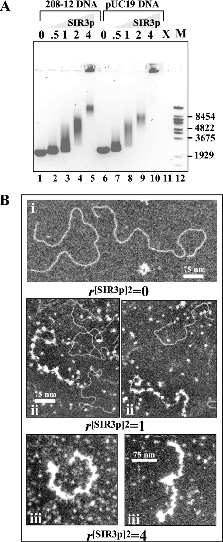
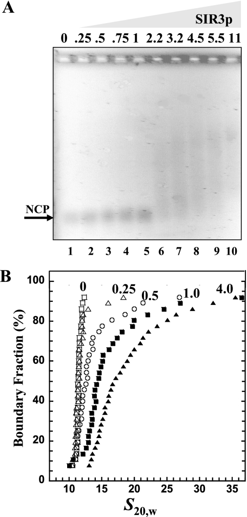
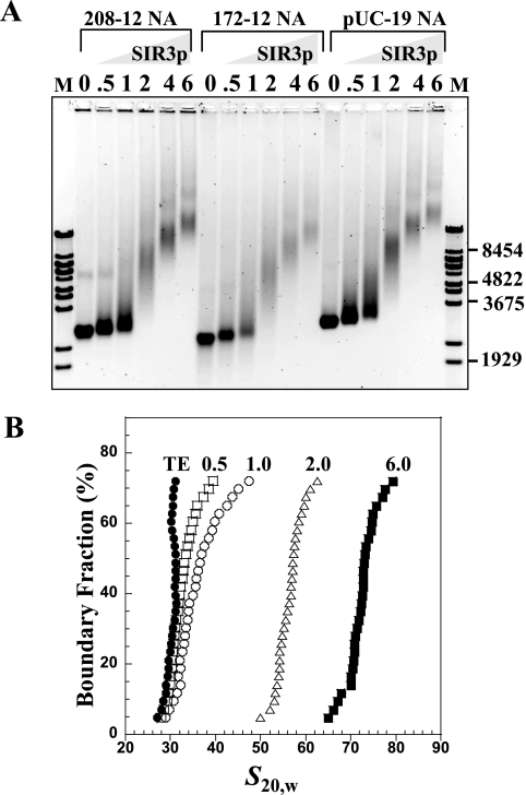
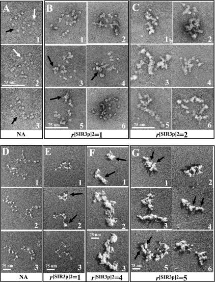
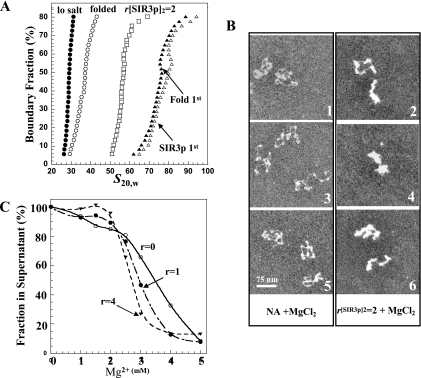
The telomeres and mating-type loci of budding yeast adopt a condensed, heterochromatin-like state through recruitment of the silent information regulator (SIR) proteins SIR2p, SIR3p, and SIR4p. In this study we characterize the chromatin binding determinants of recombinant SIR3p and identify how SIR3p mediates chromatin fiber condensation in vitro. Purified full-length SIR3p was incubated with naked DNA, nucleosome core particles, or defined nucleosomal arrays, and the resulting complexes were analyzed by electrophoretic shift assays, sedimentation velocity, and electron microscopy. SIR3p bound avidly to all three types of templates. SIR3p loading onto its nucleosomal sites in chromatin produced thickened condensed fibers that retained a beaded morphology. At higher SIR3p concentrations, individual nucleosomal arrays formed oligomeric suprastructures bridged by SIR3p oligomers. When condensed SIR3p-bound chromatin fibers were incubated in Mg(2+), they folded and oligomerized even further to produce hypercondensed higher-order chromatin structures. Collectively, these results define how SIR3p may function as a chromatin architectural protein and provide new insight into the interplay between endogenous and protein-mediated chromatin fiber condensation pathways.


Figures

FIG. 1.
FIG. 1.
Interaction of SIR3p with DNA. (A) EMSA of SIR3p and DNA. Linear 208-12 DNA (lanes 1 to 5) or linear pUC19 DNA (lanes 6 to 10) (0.5 μg; 3.6 pmol) was incubated at r[SIR3p]2 of 0.5, 1, 2, and 4 (90, 180, 360, and 720 nM SIR3p dimer, respectively) prior to electrophoresis. The positions of the markers (M, λ BstEII digest fragments) are shown at right. Lane 11 was not loaded (X). (B) EM of the interaction of SIR3p with 208-12 DNA. (i) Appearance of DNA alone after positive staining. (ii) 208-12 DNA with r[SIR3p]2 of 1 dimer per 208 unit. Some DNA molecules are studded with bound SIR3p dimers, while others have little or none. Unbound dimers are seen in the background. (iii) At r[SIR3p]2 of 4, most DNA molecules are coated with protein, and the complexes have a significantly shorter contour length. Some (apparently) free DNA is also seen. Scale bars represent 75 nm: the higher-magnification images show greater detail within the complexes.
FIG. 2.
FIG. 2.
Binding of SIR3p to NCPs. (A) EMSA. NCP 200 ng (1 pmol) of 146-bp NCPs was incubated with r[SIR3p]2 = 0, 0.25, 0.5, 0.75, 1, 2.25, 3.25, 4.5, 5.5, and 11 (12.5 to 550 nM SIR3p dimer, respectively) prior to electrophoresis on a 1% agarose gel. The gel was stained with ethidium bromide and visualized by UV transillumination. The arrow indicates the position of unbound nucleosomes. (B) Sedimentation velocity. A total of 10 μg (50 pmol) of NCP was incubated alone (□) or with r[SIR3p]2 of 0.25 (▵), 0.5(○), 1 (▪), or 4 (▴) (31, 62.5, 125, and 500 nM SIR3p dimer, respectively) prior to sedimentation velocity. The resulting integral distribution of S [corrected for water at 20°C (s20,w)] is shown.
FIG. 3.
FIG. 3.
Binding of SIR3p to nucleosomal arrays. (A) EMSA. A chromatin array with 0.5 μg (3.6 pmol) of 208-12 (lanes 2 to 7), a 172-12 chromatin array (lanes 8 to 13), and linear, assembled pUC19 DNA chromatin arrays (lanes 14 to 19) were incubated at r[SIR3p]2 of 0.5, 1, 2, 4, and 6 (per DNA repeat or per 208 bp for pUC19, net SIR3p dimer concentrations of 90, 180, 360, 720, and 1,080 nM, respectively) prior to electrophoresis on a 1% agarose gel. The gel was stained with ethidium bromide and visualized by UV transillumination. The positions of the markers (M, λ Bst-E II digest fragments) are shown at the right. (B) Sedimentation velocity. A total of 10 μg (73 pmol) of chromatin array was incubated with r[SIR3p]2 of 0 (•), 0.5 (□), 1 (○), 2 (▵), and 6 (▪) of SIR3p dimers (net SIR3p dimer concentrations of 91, 182, 365, 720, and 1,095 nM, respectively) prior to sedimentation velocity. The resulting integral distribution of S [corrected for water at 20°C (s20,w)] over the bottom 75% of the boundary is shown (the top 25% of the boundary consisted of unproductive aggregates).
FIG. 4.
FIG. 4.
EM of SIR3p-chromatin fiber complexes. (A) Negatively stained 208-12 nucleosomal arrays show 11 to 12 separated nucleosomes with linker DNA occasionally visible (arrows). (B) At r[SIR3p]2 of 1, most nucleosomes appear larger, and the total number of particles averages 17 per array, suggesting that the complexes consist of nucleosomes bound with SIR3p and SIR3p bound to linker DNA. (C) At r[SIR3p]2 of 2, arrays are coated with protein, and individual nucleosomes no longer resolved. (D to F) Glycerol dried, shadow-contrasted NAs. (D) Nucleosomes and linker DNA are clearly seen in untreated NAs. (E) At r[SIR3p]2 of 1, the population includes some that appear unchanged (E3), some that are partially compacted with fewer than 12 separate particles resolved (D1 and 2), and some that are completely compacted (D2). (F) At r[SIR3p]2 of 4, NAs appear as thickened, compact complexes (arrows) which tend to self-associate. (G) At r[SIR3p]2 of 5, thickening and self-association of arrays continues, and here arrows denote putative domains representing individual NAs. Note that with the glycerol drying technique, unbound SIR3p is separated from NAs. Scale bars represent 75 nm.
FIG. 5.
FIG. 5.
Further condensation of SIR3p-containing chromatin upon addition of MgCl2. (A) Sedimentation velocity. A total of 10 μg of nucleosomal arrays (•) was incubated with 2 mM MgCl2 for 30 min (“folded” [○]) or with SIR3p at r[SIR3p]2 of 2 for 30 min (□). The nucleosomal arrays in salt were then incubated at r[SIR3p]2 of 2 for 30 min (▵), while the chromatin fibers with r[SIR3p]2 of 2 were incubated in 2 mM MgCl2 for 30 min (▴). The resulting integral distribution of S (corrected for water at 20°C) over the bottom 80% of the boundary is shown (the top 20% of the boundary consisted of unproductive aggregates). (B) EM. Negatively stained preparations (1, 3, and 5) of nucleosomal arrays fixed in 2 mM MgCl2 are partially condensed (compare to Fig. 4D4 to 6). In the presence of r[SIR3p]2 of 2, additional condensation occurs (2, 4, and 6). Scale bars represent 100 nm. (C) Self-association of SIR3p-bound chromatin arrays. Nucleosomal arrays were incubated with r[SIR3p]2 of 0 (○), 1 (•), and 4 (▾) prior to the addition of MgCl2. The differential sedimentation assay was performed as described in Materials and Methods.

References

    1. Adams, V. H., S. J. McBryant, P. H. Wade, C. Woodcock, and J. C. Hansen. 2007. Intrinsic disorder and autonomous domain function in the multifunctional nuclear protein, MeCP2. J. Biol. Chem. 28215057-15064. - PubMed
    1. Adkins, N. L., M. Watts, and P. T. Georgel. 2004. To the 30-nm chromatin fiber and beyond. Biochim. Biophys. Acta 167712-23. - PubMed
    1. Ausio, J., D. Seger, and H. Eisenberg. 1984. Nucleosome core particle stability and conformational change: effect of temperature, particle and NaCl concentrations, and cross-linking of histone H3 sulfhydryl groups. J. Mol. Biol. 17677-104. - PubMed
    1. Barbera, A. J., J. V. Chodaparambil, B. Kelley-Clarke, V. Joukov, J. C. Walter, K. Luger, and K. M. Kaye. 2006. The nucleosomal surface as a docking station for Kaposi's sarcoma herpesvirus LANA. Science 311856-861. - PubMed
    1. Bednar, J., R. A. Horowitz, S. A. Grigoryev, L. M. Carruthers, J. C. Hansen, A. J. Koster, and C. L. Woodcock. 1998. Nucleosomes, linker DNA, and linker histone form a unique structural motif that directs the higher-order folding and compaction of chromatin. Proc. Natl. Acad. Sci. USA 9514173-14178. - PMC - PubMed

Publication types

MeSH terms

LinkOut - more resources