. 2008 May;28(10):3446-56.
doi: 10.1128/MCB.02246-07. Epub 2008 Mar 24.

p23/Sba1p protects against Hsp90 inhibitors independently of its intrinsic chaperone activity

Affiliations

p23/Sba1p protects against Hsp90 inhibitors independently of its intrinsic chaperone activity

Fedor Forafonov et al. Mol Cell Biol. 2008 May.

Abstract

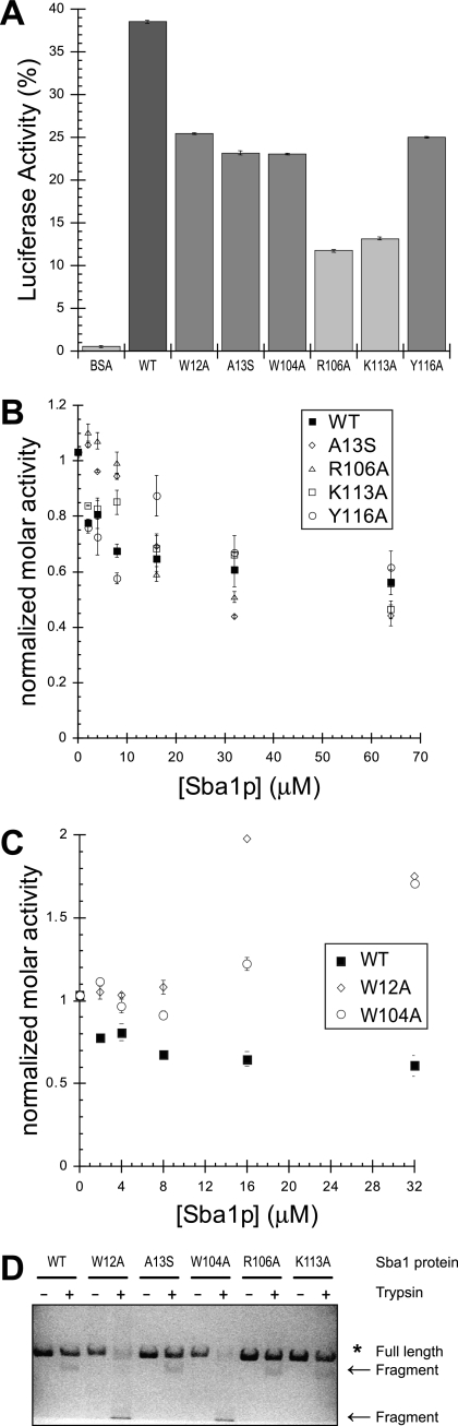
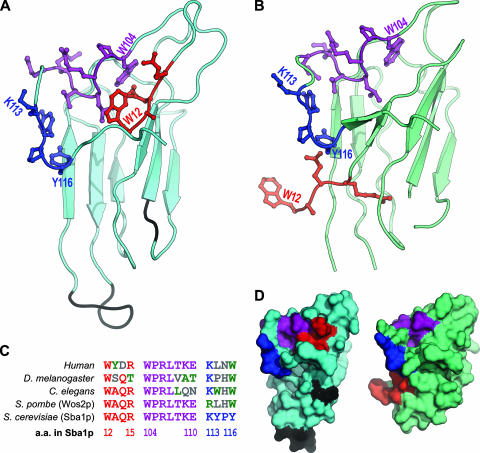
The molecular chaperone Hsp90 assists a subset of cellular proteins and is essential in eukaryotes. A cohort of cochaperones contributes to and regulates the multicomponent Hsp90 machine. Unlike the biochemical activities of the cochaperone p23, its in vivo functions and the structure-function relationship remain poorly understood, even in the genetically tractable model organism Saccharomyces cerevisiae. The SBA1 gene that encodes the p23 ortholog in this species is not an essential gene. We found that in the absence of p23/Sba1p, yeast and mammalian cells are hypersensitive to Hsp90 inhibitors. This protective function of Sba1p depends on its abilities to bind Hsp90 and to block the Hsp90 ATPase and inhibitor binding. In contrast, the protective function of Sba1p does not require the Hsp90-independent molecular chaperone activity of Sba1p. The structure-function analysis suggests that Sba1p undergoes considerable structural rearrangements upon binding Hsp90 and that the large size of the p23/Sba1p-Hsp90 interaction surface facilitates maintenance of high affinity despite sequence divergence during evolution. The large interface may also contribute to preserving a protective function in an environment in which Hsp90 inhibitory compounds can be produced by various microorganisms.


Figures

FIG. 1.
FIG. 1.
Δsba1 strains are hypersensitive to radicicol (Rd). Shown is a growth assay with 10-fold serial dilutions of cells of the four isogenic strains with the indicated genotypes.
FIG. 2.
FIG. 2.
p23-negative MEFs are hypersensitive to GA. (A) Growth curves for wild-type (WT) or p23-null (−/−) cells cultured without or with 40 nM GA (added at day 0). (B) Ratios of numbers of treated over untreated cells. Cell numbers for each experimental series were standardized to the experimentally determined values on day 0 (set to 100%). Error bars show standard deviations (for clarity only on one side of data points).
FIG. 3.
FIG. 3.
Hsp90 interaction and in vivo complementation of full-length and truncated forms of Sba1p. (A) Schematic representation of the Sba1p truncation mutants. The N-terminal Flag tag and removed residues are indicated. The p23/Sba1 signature motif is underlined. (B) Sba1p interacts with both Hsp82p and Hsc82p. An immunoblot of an Hsp90 co-IP experiment is shown. Flag-tagged Sba1p was expressed in isogenic strains expressing either one or both Hsp90 isoforms and used to co-IP Hsp90 with an anti-Flag resin. The immunoblot was probed with polyclonal Hsp90 antibodies recognizing both isoforms (see lanes labeled “input”; the uppermost bands correspond to full-length Hsp82p and Hsc82p, whereas the lower bands are degradation products). (C) Expression and Hsp90 interaction of Sba1p truncation mutants. Flag-tagged Sba1p derivatives shown in panel A were expressed in the Δsba1 strain. Total extracts (40 μg each) and anti-Flag immunoprecipitates were immunoblotted with anti-Flag and anti-Hsp90 antibodies as indicated. In the case of the full-length Flag-Sba1b and the Δ131-216 mutant, five times less immunoprecipitate was loaded to facilitate the comparison with the more poorly expressed mutants. (D) The 130 N-terminal amino acids of Sba1p are sufficient for conferring radicicol (Rd) resistance. A growth assay with fivefold serial dilutions of Δsba1 Δpdr5 cells expressing the Sba1p proteins indicated on the left was performed with increasing concentrations of radicicol or with GA at 30 and 37°C.
FIG. 4.
FIG. 4.
Hsp90 interaction and in vivo complementation of Sba1p point mutants. Both wild-type Sba1p (WT) and point mutants were expressed as Flag-tagged versions in the Δsba1 Δpdr5 strain. (A) Expression and Hsp90 interaction of Sba1p point mutants. The immunoblots represent two different co-IP experiments. ATP was present during the incubation with the anti-Flag resin for both experiments, but in co-IP experiment 1 (IP1), ATP was omitted from the buffer used to wash the immunoprecipitates. Note that a doublet is observed for Flag-Sba1 in some experiments, presumably because of C-terminal cleavage. The weaker band below Flag-Sba1 is due to the immunoglobulin light chain. For other details, see Materials and Methods and the legend to Fig. 3C. (B) Radicicol (Rd) sensitivity of the Δsba1 Δpdr5 strains expressing Sba1p point mutants. Growth assays were done as described in the legend to Fig. 3D.
FIG. 5.
FIG. 5.
In vitro characterization of biochemical activities of Sba1p point mutants. (A) Sba1p point mutants retain molecular chaperone activity. The bar graph shows recovered luciferase activities, which are indicative of the ability of wild-type Sba1p (WT) and its point mutants to maintain denatured firefly luciferase in a refoldable state for refolding by Hsp70 and Hdj1. BSA was used as a negative control. The data represent the average values from three independent experiments. (B and C) The Sba1p point mutants display differential effects on ATP hydrolysis by Hsp82p. ATPase activity is expressed as a function of Sba1p concentrations. The data for the five Sba1p mutants are presented in two separate graphs to facilitate reading and represent the average values from three independent experiments. (D) The Sba1p mutants that do not inhibit the ATPase of Hsp82p have distinct conformational states. The structures of Sba1p mutants were probed using limited proteolytic digestion with trypsin. After digestion for 0 (lanes marked “−”) and 15 min (lanes marked “+”), samples were resolved by sodium dodecyl sulfate-polyacrylamide gel electrophoresis and visualized by Coomassie blue staining.
FIG. 6.
FIG. 6.
Sba1p reduces binding affinity of Hsp90 for GA. The graph displays the change in the fluorescence anisotropy of fluorescein-labeled GA (FITC-GA) that it undergoes as it binds Hsp82p. The data points (averages of four replicates) and fitted curves indicate the changes in the absence or presence of a fixed amount of wild-type (WT) or representative mutant Sba1p proteins. The Kds calculated from the fitted curves are shown within the legend. Curves for additional mutants were omitted for clarity. Their Kds for A13S, R106A, and L107S are 60.8 ± 6.3, 51.1 ± 6.7, and 23.6 ± 4.0 nM, respectively.
FIG. 7.
FIG. 7.
The Y116A-Hsp90 interaction is insensitive to radicicol (Rd). Shown are immunoblots of a co-IP experiment with antibodies to Flag-tagged wild-type (WT) or mutant (Y116A) Sba1p expressed in a Δsba1 yeast strain. A fraction of the crude yeast extract was loaded as “input,” whereas the anti-Flag immunoprecipitate was either analyzed directly or after further incubation as indicated.
FIG. 8.
FIG. 8.
p23 protects the Hsp90 substrate Raf-1 against GA-induced degradation. Shown is a representative immunoblot of extracts from wild-type (WT) and p23-null MEFs treated with 60 nM GA for the indicated times. Raf-1 levels were standardized to GAPDH, and the starting level was set to 100% (see numbers below the micrograph).
FIG. 9.
FIG. 9.
Model of the structural rearrangements of Sba1p induced by Hsp90 binding. (A) Model of the crystal structure of the N-terminal domain of Sba1p (residues 12 to 121). To model the Sba1p sequence over the known structure of the unbound human p23 (60), we generated a multiple-sequence alignment and submitted it to the fully automated protein structure homology-modeling server SWISS-MODEL (55). Residues 1 to 11 were omitted because they are not resolved in the published structure of the Sba1p-Hsp82p complex (2). (B) Experimental crystal structure of Sba1p in a complex with Hsp82p (2). Rendering of the Sba1p structure from PDB file 2CG9 was done with the PyMOL molecular graphics system (W. L. DeLano, 2002; http://www.pymol.org). (C) Sequence alignment of the mutagenized sequence motifs. Colors other than red, purple, and blue are used for residues that are not conserved, with green and gray being the most and the least similar ones, respectively. (D) Surface representations of the two structures shown in panels A and B. Black portions in the model for unbound Sba1p correspond to two loops that are not resolved in the published structure of the complex.

Similar articles

Cited by

References

    1. Abbas-Terki, T., O. Donzé, P.-A. Briand, and D. Picard. 2001. Hsp104 interacts with Hsp90 cochaperones in respiring yeast. Mol. Cell. Biol. 217569-7575. - PMC - PubMed
    1. Ali, M. M., S. M. Roe, C. K. Vaughan, P. Meyer, B. Panaretou, P. W. Piper, C. Prodromou, and L. H. Pearl. 2006. Crystal structure of an Hsp90-nucleotide-p23/Sba1 closed chaperone complex. Nature 4401013-1017. - PMC - PubMed
    1. Bandhakavi, S., R. O. McCann, D. E. Hanna, and C. V. Glover. 2003. A positive feedback loop between protein kinase CKII and Cdc37 promotes the activity of multiple protein kinases. J. Biol. Chem. 2782829-2836. - PubMed
    1. Bohen, S. P. 1998. Genetic and biochemical analysis of p23 and ansamycin antibiotics in the function of Hsp90-dependent signaling proteins. Mol. Cell. Biol. 183330-3339. - PMC - PubMed
    1. Bose, S., T. Weikl, H. Bügl, and J. Buchner. 1996. Chaperone function of Hsp90-associated proteins. Science 2741715-1717. - PubMed

Publication types

MeSH terms