. 2008 Mar 24;180(6):1115-31.
doi: 10.1083/jcb.200708170.

Dissection of the essential steps for condensin accumulation at kinetochores and rDNAs during fission yeast mitosis

Affiliations

Dissection of the essential steps for condensin accumulation at kinetochores and rDNAs during fission yeast mitosis

Norihiko Nakazawa et al. J Cell Biol. .

Abstract

The condensin complex has a fundamental role in chromosome dynamics. In this study, we report that accumulation of Schizosaccharomyces pombe condensin at mitotic kinetochores and ribosomal DNAs (rDNAs) occurs in multiple steps and is necessary for normal segregation of the sister kinetochores and rDNAs. Nuclear entry of condensin at the onset of mitosis requires Cut15/importin alpha and Cdc2 phosphorylation. Ark1/aurora and Cut17/Bir1/survivin are needed to dock the condensin at both the kinetochores and rDNAs. Furthermore, proteins that are necessary to form the chromatin architecture of the kinetochores (Mis6, Cnp1, and Mis13) and rDNAs (Nuc1 and Acr1) are required for condensin to accumulate specifically at these sites. Acr1 (accumulation of condensin at rDNA 1) is an rDNA upstream sequence binding protein that physically interacts with Rrn5, Rrn11, Rrn7, and Spp27 and is required for the proper accumulation of Nuc1 at rDNAs. The mechanism of condensin accumulation at the kinetochores may be conserved, as human condensin II fails to accumulate at kinetochores in hMis6 RNA interference-treated cells.


Figures

Figure 1.
Figure 1.
Mitotic nuclear accumulation of condensin and its loss in two mutants. (A) Time-lapse images of the wild-type S. pombe cultured at 36°C. Cells expressed the condensin subunit Cnd1-GFP (green) and the SPB and nuclear envelope protein Sad1-RFP (red). In the inset, the arrowheads indicate the two enriched nuclear domains. The arrow in the merged images shows the condensin in the middle of the two separated daughter nuclei. (bottom) The mitotic nucleus containing Cut14-GFP and stained with the ethidium bromide–DAPI mixture, which revealed the nucleolar domain in the mitotic nucleus. (B and C) Two example cells in metaphase at 1-min intervals are shown that expressed Cnd1-GFP Sad1-RFP (B) or Cut14-GFP Sad1-RFP (C). (D) Time-lapse images for a wild-type cell carrying plasmid REP81-Cut3WT (left) or REP81-Cut3 T19A (right). (E and F) Time-lapse images of mutant cut15-85 that expressed Cut14-GFP (E) or Cnd1-GFP (F) and Sad1-mCherry. (A and D–F) The numbers in the panels indicate time in minutes. Bars: (A, top; and B–F) 10 μm; (A, bottom) 2 μm.
Figure 2.
Figure 2.
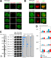
Cnp1, Mis6, and Mis13 are required for localizing condensin at the kinetochore. (A) Cells cultured at 26°C in EMM2 were observed for the colocalization of Cnd1-GFP with Mis12-RFP, a centromere/kinetochore protein. The numbers in the right panels indicate time in minutes. The enlarged images of Cnd1-GFP (left), Mis12-RFP (middle), and the merged images (right) at 13 min are shown in the insets. (B) Cut14-GFP and Sad1-RFP were observed in the wild-type, mis6-302, cnp1-1, mis13-1, mis16-53, and mis18-262 mutants cultured at 26°C and were shifted to 36°C for 8 h. (C) Chromosomally integrated Cnp1/CENP-A–GFP expressed under the native promoter was observed in the wild-type and cut14-208 mutant cultured at 36°C for 2 h. (D) A ChIP assay was performed using extracts of block-released nda3-311 mutant that expressed Cut14-Flag. The probes were from the central centromere, cnt1 (c10, c9, and c7.5), imr1 (c4 and c1), the outer centromere dg, and the noncentromeric lys1 +. WCE, whole cell extract; IP, immunoprecipitate. Quantitative data are shown at the bottom (see Results for details). (E) A ChIP assay was performed using extracts of block-released cdc25-22 mutant that expressed Cut14-Flag. Extracts of cdc25 cells (blocked in G2 at 36°C and released to 26°C) were prepared at 0 min (G2 phase), 25 min (prometaphase), 50 min (meta/anaphase), and 75 min (telophase or G1/S phase). The probes used were from the central centromere, cnt1 (c9), imr1 (c4), and the three arm probes arm1 (SPBC28F2.08c coding region), arm2 (SPBC29B5 noncoding region), and arm3 (ars2004). In the bottom panel, the percent frequencies of different types of cells are indicated.(F) A ChIP assay was performed using extracts of asynchronous wild-type, mis6-302, and cnp1-1 mutants that expressed Cut14-Flag. These strains were cultured at 26°C and were shifted to 36°C for 8 h. Real-time PCR was performed using the primers of cnt1, imr1, lys1 +, and rDNA (N1). The levels of precipitated cnt1 and imr1 were diminished in cnp1-1 and mis6-302 mutants. Because the asynchronous cultures were used, the differences between wild-type and mutant cells were relatively small. Error bars represent SD. Bars: (A and C) 10 μm; (B) 2 μm.
Figure 3.
Figure 3.
RNAi of hMis6 diminishes condensin II at human kinetochores. (A and B) Kinetochore localization of hCAP-H2 on the spread metaphase chromosomes is diminished in cells after RNAi knockdown. Metaphase spread chromosomes were prepared in HeLa-transfected cells for 48 h by siRNA of hMis6 (A) or luciferase (B; control). Cells were stained by Hoechst33342 for DNA (blue), anti–CENP-A antibody for the centromere/kinetochore (red), and anti–hCAP-H2 for the condensin (green) antibodies. The enlarged images correspond to the dashed boxes in the left panel. (C) The percent frequencies of metaphase chromosomes showing the hCAP-H2 signals on kinetochores; the control was 98.2% (n = 112), whereas depleted hMis6 was only 18.3% (n = 131). (D) HeLa transfected by siRNA of luciferase or hMis6 were fixed for 48 h and stained by Hoechst33342 (blue), anti-hMis6 (red), and anti–hCAP-H2 (green) antibodies. Bars, 10 μm.
Figure 4.
Figure 4.
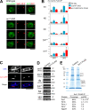
Nuc1 is required for localizing condensin at rDNAs. (A) Localization of Cut14-GFP was examined in the nuc1-632 mutant cultured at 36°C for 6 h. (B) Localization of Cut14-GFP was examined in the nuc1-632 mutant with the nucleolar protein Gar2-mCherry. (C) A ChIP assay was performed using a block-release nda3-311 mutant that expressed Cut14-Flag. Nine DNA probes derived from the rDNA repeat unit were used: NTS (N1–N4), ARS, external transcribed sequence, and 18S, 5.8S, and 28S coding sequences. WCE, whole cell extract; IP, immunoprecipitate. The negative control (−) is the strain that did not carry Cut14-Flag. Quantitative enrichment is shown on the right. (D) A ChIP assay was performed using extracts of asynchronous wild-type and nuc1-632 mutant that expressed Cut14-Flag. The wild-type and nuc1-632 mutant were cultured at 26°C and shifted to 36°C for 6 h, and immunoprecipitation was performed. Real-time PCR was used to amplify and quantify DNAs with the PCR primers (N1, N3, lys1 +, and cnt1). Error bars represent SD. Bars, 2 μm.
Figure 5.
Figure 5.
Enrichment of condensin at rDNAs requires a nucleolar protein, Acr1, which interacts with the rDNA upstream activator complex. (A) Cut14-GFP was observed in mitotic cells of the acr1-936 mutant and the control wild type cultured at 36°C for 4 h. (B) A ChIP assay was performed using extracts of asynchronous wild-type and acr1-936 mutant that expressed Cut14-Flag. The wild-type and acr1-936 mutant were cultured at 26°C and shifted to 36°C for 4 h, and immunoprecipitation was performed. Real-time PCR was used to amplify and quantify DNAs with the PCR primers (N1, N2, N3, lys1 +, and cnt1). Error bars represent SD. (C) Localization of Acr1 was examined in the strain that expressed Acr1-GFP. (D) A ChIP assay was performed using the strain that expressed Acr1-Flag. Two rDNA probes (N2 and 18S) and four control probes (cnt1, imr1, dg, and lys1 +) were used. (E, top) Preparation of proteins coprecipitated with Acr1-Flag and the Flag-only control for liquid chromatography–tandem mass spectrometry. The positions of identified protein names are indicated. The two bands were obtained for Acr1-Flag (Fig. S2 D, available at http://www.jcb.org/cgi/content/full/jcb.200708170/DC1). (bottom) Four proteins (Rrn5, Rrn11, Rrn7, and Spp27) were coprecipitated with Acr1-Flag. emPAI, exponentially modified protein abundance index (Ishihama et al., 2005). Bars: (A) 2 μm; (C) 10 μm.
Figure 6.
Figure 6.
acr1-936 had both mitotic and interphase defects. (A) DAPI-stained wild-type, nuc1-632, and acr1-936 cells that were cultured at 36°C for 4–8 h. The insets correspond to the white dashed boxes in the left panels. (B) Localization of Nuc1 was dispersed in the nucleus in the acr1-936 mutant. Bars, 10 μm.
Figure 7.
Figure 7.
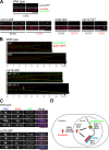
Condensin entered the nucleus but failed to accumulate at the two distinct domains in the cut17 mutant and the dominant-negative Ark1/aurora cells. (A, top) Wild-type and cut17-275 mutant cells that expressed Cut14-GFP and Sad1-mCherry were cultured at 26°C, shifted to 36°C for 3 h, and observed. (bottom) The wild-type cells carrying the inducible plasmid REP1-Ark1WT or REP1-Ark1-K118R were cultured in the absence of thiamine at 26°C. (B) Time-lapse series of the Cut14-GFP and Sad1-mCherry images in the wild-type or cut17-275 cells cultured at 36°C for 3 h (top) and in the cells expressing wild-type Ark1 or the dominant-negative Ark1 K118R are shown. Bars, 10 μm.
Figure 8.
Figure 8.
Proper segregation of sister kinetochores and rDNAs requires condensin. (A) Cen2-GFP was used to examine the segregation phenotype of centromeric DNA in the condensin mutant cut14-208. The wild type, mis6-302, and mis12-537 were used as the control strains. These were first grown at 26°C and were shifted to 36°C for 2 h (wild type and cut14-208) or 8 h (mis6-302 and mis12-537). The frequency of equal or unequal segregation is shown at the bottom of the images. (B) Kymographs for Cen2-GFP are shown for the wild type and cut14-208, which also expressed Sad1-RFP for the SPB marker. Images were taken at 10-s intervals. The frequency of each phenotype is indicated in parentheses. The arrow in the bottom panel (cell 2) shows the two closely situated splits of Cen2-GFP signals upon the presumed onset of anaphase. (C) The rDNA repeat unit was used for the FISH probe in the wild-type and condensin mutant cells. DAPI and anti-Sad1 antibodies were used to stain DNA and to immunostain the SPBs, respectively. (D) Steps required for condensin to be accumulated at mitotic kinetochores and rDNAs in S. pombe. Cut15, importin α; Cut17, Bir1/survivin; Ark1, aurora; Cnp1, CENP-A; Mis6, CENP-I; Mis13, hMis13; Nuc1, pol I largest subunit; Acr1, accumulation of condensin at rDNA. Bars: (A) 5 μm; (B), 2 μm; (C) 10 μm.

Similar articles

Cited by

References

    1. Aoki, K., Y. Nakaseko, K. Kinoshita, G. Goshima, and M. Yanagida. 2006. CDC2 phosphorylation of the fission yeast dis1 ensures accurate chromosome segregation. Curr. Biol. 16:1627–1635. - PubMed
    1. Aono, N., T. Sutani, T. Tomonaga, S. Mochida, and M. Yanagida. 2002. Cnd2 has dual roles in mitotic condensation and interphase. Nature. 417:197–202. - PubMed
    1. Appelgren, H., B. Kniola, and K. Ekwall. 2003. Distinct centromere domain structures with separate functions demonstrated in live fission yeast cells. J. Cell Sci. 116:4035–4042. - PubMed
    1. Bhalla, N., S. Biggins, and A.W. Murray. 2002. Mutation of YCS4, a budding yeast condensin subunit, affects mitotic and nonmitotic chromosome behavior. Mol. Biol. Cell. 13:632–645. - PMC - PubMed
    1. Cabello, O.A., E. Eliseeva, W.G. He, H. Youssoufian, S.E. Plon, B.R. Brinkley, and J.W. Belmont. 2001. Cell cycle-dependent expression and nucleolar localization of hCAP-H. Mol. Biol. Cell. 12:3527–3537. - PMC - PubMed

Publication types

MeSH terms