. 2008 Aug;22(8):2629-38.
doi: 10.1096/fj.08-107169. Epub 2008 Mar 24.

Apoptosis induces expression of sphingosine kinase 1 to release sphingosine-1-phosphate as a "come-and-get-me" signal

Affiliations

Apoptosis induces expression of sphingosine kinase 1 to release sphingosine-1-phosphate as a "come-and-get-me" signal

David R Gude et al. FASEB J. 2008 Aug.

Abstract

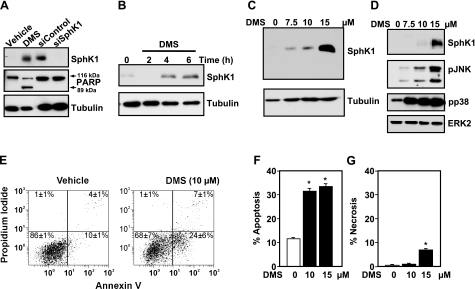
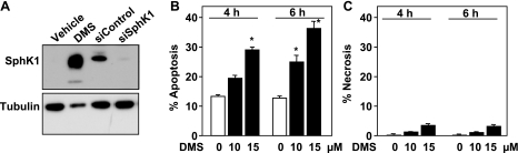
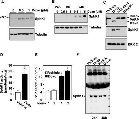
Sphingosine-1-phosphate (S1P) is a bioactive lipid that regulates myriad important cellular processes, including growth, survival, cytoskeleton rearrangements, motility, and immunity. Here we report that treatment of Jurkat and U937 leukemia cells with the pan-sphingosine kinase (SphK) inhibitor N,N-dimethylsphingosine to block S1P formation surprisingly caused a large increase in expression of SphK1 concomitant with induction of apoptosis. Another SphK inhibitor, D,L-threo-dihydrosphingosine, also induced apoptosis and produced dramatic increases in SphK1 expression. However, up-regulation of SphK1 was not a specific effect of its inhibition but rather was a consequence of apoptotic stress. The chemotherapeutic drug doxorubicin, a potent inducer of apoptosis in these cells, also stimulated SphK1 expression and activity and promoted S1P secretion. The caspase inhibitor ZVAD reduced not only doxorubicin-induced lethality but also the increased expression of SphK1 and secretion of S1P. Apoptotic cells secrete chemotactic factors to attract phagocytic cells, and we found that S1P potently stimulated chemotaxis of monocytic THP-1 and U937 cells and primary monocytes and macrophages. Collectively, our data suggest that apoptotic cells may up-regulate SphK1 to produce and secrete S1P that serves as a "come-and-get-me" signal for scavenger cells to engulf them in order to prevent necrosis.


Figures

Figure 1.
Figure 1.
SphK inhibitor DMS induces SphK1 expression. A–D) Jurkat T cells were treated without or with 15 μM DMS for 6 h (A) or the indicated time (B) or with the indicated concentration of DMS for 8 h (C) or 4 h (D). Equal amounts of cell lysates (20 μg) were separated by SDS-PAGE and immunoblotted with anti-SphK1 antibody. Blots were stripped and reprobed with anti-tubulin antibody (A–C) or with anti-extracellular regulated kinase (ERK) 2 (D) as loading controls. Blots were probed with anti-PARP antibody as a marker of apoptosis (A); arrows indicate p116 full-length PARP and its p89 fragment; lysates (20 μg) from M2 melanoma cells transfected with scrambled siRNA or siRNA targeted to SphK1 were included as controls to indicate the molecular weight of SphK1. Cell lysates were also immunoblotted with pJNK and pp38 antibodies (D). E–G) Jurkat T cells were treated with vehicle or the indicated concentrations of DMS for 6 h, then stained with annexin V/PI, and apoptosis was determined by flow cytometry, as described in Materials and Methods. The percentages in the bottom right quadrants (E) correspond to early apoptotic cells (annexin V-positive), whereas percentages in the top right quadrants correspond to late apoptotic cells (annexin V- and PI-positive). Apoptotic cells are annexin V-positive (F), whereas necrotic cells are PI-positive only (G). Similar results were obtained in four independent experiments. *P < 0.01.
Figure 2.
Figure 2.
DMS increases expression of SphK1 in U937 cells. U937 cells were treated with vehicle or with 15 μM DMS for 24 h (A) or with the indicated concentrations of DMS for 4 or 6 h (B, C). A) Cells were lysed and equal amounts of protein (60 μg) were separated by SDS-PAGE and immunoblotted with anti-SphK1 antibody. Blots were stripped and reprobed with anti-tubulin antibody as loading controls. Lysates from M2 melanoma cells transfected with scrambled siRNA or siRNA targeted to SphK1 were included as controls to indicate the molecular weight of SphK1. B, C) Cells were stained with annexin V/PI. Apoptotic cells are annexin V-positive (B), whereas necrotic cells are PI-positive only (C). Similar results were obtained in four independent experiments. *P < 0.05.
Figure 3.
Figure 3.
Another SphK inhibitor, DHS, increases expression of SphK1 and induces apoptosis in Jurkat cells. A) Jurkat cells were treated for 18 h without or with 10 or 20 μM DHS. Equal amounts of cell lysates were separated by SDS-PAGE and immunoblotted with anti-SphK1 antibody. Blots were stripped and reprobed with anti-tubulin antibody as loading controls. B) Cell lysates (50 μg) from DHS-treated Jurkat cells were immunoprecipitated with either anti-SphK1 antibody or rabbit IgG. Equal amounts of each immunoprecipitate were separated by SDS-PAGE and immunoblotted with anti-SphK1 antibody. C–F) Jurkat cells were treated for 6 h without or with the indicated concentrations of DHS, then stained with annexin V/PI. Apoptotic cells are annexin V-positive (C), whereas necrotic cells are PI-positive only (D). Representative results are shown in E and F. The percentages in the bottom right quadrants correspond to early apoptotic cells (annexin V-positive), whereas percentages in the top right quadrants correspond to late apoptotic cells (annexin V- and PI-positive). Similar results were obtained in four independent experiments. *P < 0.01.
Figure 4.
Figure 4.
Doxorubicin increases SphK1 expression, S1P secretion, and apoptosis. Jurkat cells were treated with the indicated concentrations of doxorubicin (A) or with 0.5 μM (C–F) for 24 h. U937 cells were treated with the indicated concentrations of doxorubicin for 8 or 24 h (B). A, B) Equal amounts of cell lysates were separated by SDS-PAGE and immunoblotted with anti-SphK1 antibody. Blots were stripped and reprobed with anti-tubulin antibody as a loading control. C) Blots were also probed with anti-PARP antibody. Arrows indicate p116 full length PARP and its p89 fragment. Blots were stripped and reprobed with anti-ERK2 as a loading control. Lysates from M2 melanoma cells transfected with scrambled siRNA or siRNA targeted to SphK1 (20 μg) were included as controls to indicate the molecular weight of SphK1. D) SphK1 activity was determined in lysates from Jurkat cells treated with vehicle or doxorubicin (0.5 μM) for 24 h. E) Jurkat cells were treated with vehicle (open bars) or doxorubicin (0.5 μM, solid bars) for 24 h, labeled for 10 min with [3H]sphingosine, and washed extensively, and [3H]S1P secretion was determined after 1 or 2 h, as described in Materials and Methods. F) Jurkat cells were treated with vehicle or 0.5 μM doxorubicin (Doxo) for 24 or 48 h. The medium was collected and concentrated, and proteins were analyzed by immunoblotting with anti-SphK1 antibody. Similar results were obtained in two independent experiments. *P < 0.01.
Figure 5.
Figure 5.
Inhibition of apoptosis suppresses doxorubicin-induced SphK1 expression and S1P secretion. A) Jurkat cells were pretreated for 15 min with 40 μM ZVAD and then treated for the indicated time with 0.5 μM doxorubicin. Equal amounts of cell lysate proteins were separated by SDS-PAGE and immunoblotted with anti-SphK1 antibody or with anti-PARP antibody. Arrows indicate p116 full length PARP and its p89 fragment. Blots were stripped and reprobed with anti-tubulin as a loading control. Lysates from cells transfected with scrambled siRNA or siRNA targeted to SphK1 (20 μg) were included as controls to indicate the molecular weight of SphK1. B–D) Jurkat cells were pretreated for 15 min with 40 μM ZVAD and then treated with 0.5 μM doxorubicin for 24 h. Cells were stained with annexin V/PI, and apoptosis (B) or necrosis (C) was determined by flow cytometry, as described in Materials and Methods. Cells (106) were labeled for 10 min with [3H]sphingosine, washed extensively, and [3H]S1P secretion was determined at 1 and 2 h (D), as described in Materials and Methods. Similar results were obtained in two independent experiments. *P < 0.01 vs. vehicle; **P < 0.01 vs. samples without ZVAD treatment.
Figure 6.
Figure 6.
S1P is a potent chemoattractant for monocytic cell lines and primary monocytes. Vehicle or S1P was added at increasing concentrations (1, 10, 100, and 1000 nM; solid bars) directly to the lower chamber, and transmigration of U937 cells (A) and THP-1 monocytic cells (B) was determined as described in Materials and Methods. CCL2 (50 ng/ml; shaded bar) was used as a positive control. Primary monocytes (C) or macrophages (D) were allowed to migrate toward vehicle, increasing concentrations of S1P (1, 10, 100, and 1000 nM; solid bars), or 10% FCS (shaded bar) in modified Boyden chambers. Data are expressed as number of migrating cells per field and are means ± sd of triplicate determinations. Similar results were obtained in two independent experiments. *P < 0.001.

References

    1. Spiegel S, Milstien S. Sphingosine-1-phosphate: an enigmatic signalling lipid. Nat Rev Mol Cell Biol. 2003;4:397–407. - PubMed
    1. Ogretmen B, Hannun Y A. Biologically active sphingolipids in cancer pathogenesis and treatment. Nat Rev Cancer. 2004;4:604–616. - PubMed
    1. Cuvillier O, Pirianov G, Kleuser B, Vanek P G, Coso O A, Gutkind S, Spiegel S. Suppression of ceramide-mediated programmed cell death by sphingosine-1-phosphate. Nature. 1996;381:800–803. - PubMed
    1. Taha T A, Hannun Y A, Obeid L M. Sphingosine kinase: biochemical and cellular regulation and role in disease. J Biochem Mol Biol. 2006;39:113–131. - PubMed
    1. Huwiler A, Pfeilschifter J. Altering the sphingosine-1-phosphate/ceramide balance: a promising approach for tumor therapy. Curr Pharm Des. 2006;12:4625–4635. - PubMed

Publication types

MeSH terms

LinkOut - more resources