. 2008 Apr 1;105(13):5028-33.
doi: 10.1073/pnas.0710576105. Epub 2008 Mar 24.

The editing enzyme ADAR1 and the mRNA surveillance protein hUpf1 interact in the cell nucleus

Affiliations

The editing enzyme ADAR1 and the mRNA surveillance protein hUpf1 interact in the cell nucleus

Lily Agranat et al. Proc Natl Acad Sci U S A. .

Abstract

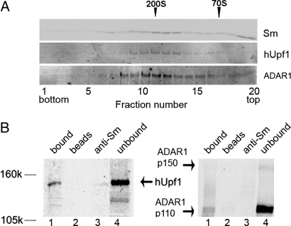
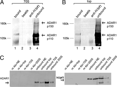
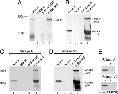
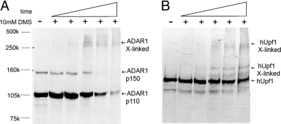
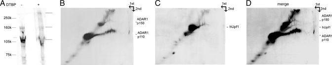
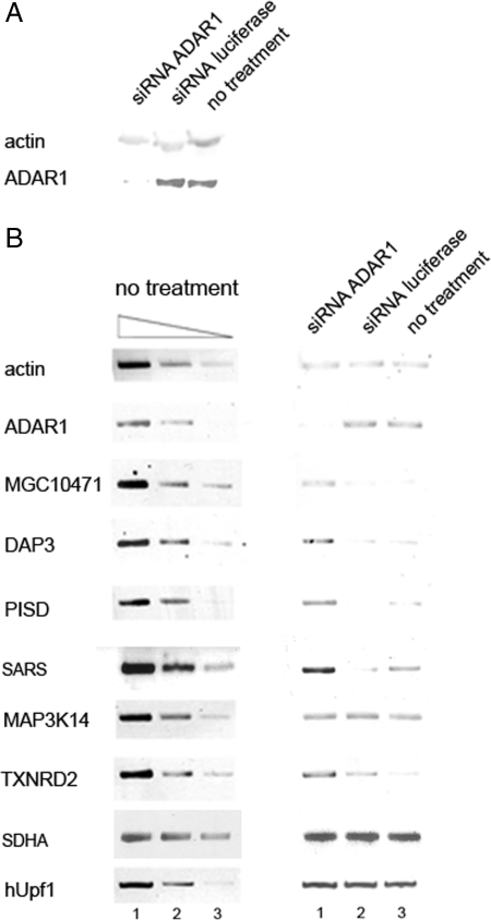
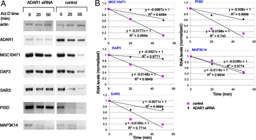
Posttranscriptional regulation is an important step in the regulation of gene expression. In this article, we show an unexpected connection between two proteins that participate in different processes of posttranscriptional regulation that ensures the production of functional mRNA molecules. Specifically, we show that the A-to-I RNA editing protein adenosine deaminase that acts on RNA 1 (ADAR1) and the human Upf1 (hUpf1) protein involved in RNA surveillance are found associated within nuclear RNA-splicing complexes. A potential functional role for this association was revealed by RNAi-mediated down-regulation of ADAR1, which was accompanied by up-regulation of a number of genes previously shown to undergo A-to-I editing in Alu repeats and to be down-regulated by hUpf1. This study suggests a regulatory pathway by a combination of ADAR1 A-to-I editing enzyme and RNA degradation presumably with the aid of hUpf1.


Conflict of interest statement

The authors declare no conflict of interest.

Figures

Fig. 1.
Fig. 1.
hUpf1 is associated with supraspliceosomes. (A) hUpf1 sediments with supraspliceosomes. Supraspliceosomes prepared from HeLa cell nuclei as described in refs. and were fractionated in a sucrose gradient. The 200S fractions were refractionated in a second sucrose gradient. Aliquots from each fraction were run on an SDS/PAGE and were Western blotted with anti-Sm, anti-hUpf1, and anti-ADAR1 antibodies. The sedimentation of 200S TMV and 70S bacterial ribosomes size markers are indicated above the top row. (B) Indirect IP of hUpf1 and ADAR1 from supraspliceosomes. Supraspliceosomes were immunoprecipitated by anti-Sm antibodies, and the precipitated and unbound proteins were analyzed by SDS/PAGE and probed by anti-hUpf1 (Left) and ADAR1 (Right) antibodies (lanes 1 and 4, respectively). As controls, we show the reaction without the antibody (lane 2) and the reaction with antibodies and buffer instead of the sample (lane 3).
Fig. 2.
Fig. 2.
hUpf1 is associated with nuclear complexes together with ADAR1. (A) Nuclear complexes sedimenting at 70S were immunoprecipitated by anti-hUpf1 antibodies, and the precipitated and unbound proteins were analyzed by SDS/PAGE and probed by anti-ADAR1 antibodies (lanes 1 and 4, respectively). Controls: lane 2, no antibody; lane 3, no sample. (B) Nuclear complexes sedimenting at the top of the gradient were immunoprecipitated as described in A (lanes in B as in A). (C) HeLa cell NE and nuclear complexes sedimenting at the top of the gradient at 70S and at 200S were immunoprecipitated by a nonrelevant antibody, preimmune anti-rabbit IgG, and were Western blotted by anti-ADAR1 (Left) and anti-hUpf1 (Right) antibodies.
Fig. 3.
Fig. 3.
ADAR1 and hUpf1 are associated in HeLa cell NE. (A) HeLa cell NE was immunoprecipitated by anti-ADAR1 antibodies, and the precipitated and unbound proteins were analyzed by SDS/PAGE and probed by anti-hUpf1 antibodies (lanes 1 and 4, respectively). Controls: lane 2, no antibody; lane 3, no sample. (B) In a complementary experiment, we show IP of HeLa cell NE by anti-hUpf1 antibodies and Western blotting by anti-ADAR1 antibodies. (C) HeLa cell NE was subjected to RNase A treatment and was then immunoprecipitated by anti-hUpf1 antibodies and Western blotted by ADAR1. The lanes correspond to those in A. (D) The same as in C, except that the NE was treated with RNase V1. (E) RT-PCR of actin RNA extracted from the HeLa cell NE treated and untreated with RNase A (Upper) and RNase V1 (Lower) shown in C and D.
Fig. 4.
Fig. 4.
Chemical cross-linking results in formation of oligomers containing ADAR1 and hUpf1. HeLa cell NE was cross-linked by DMS for increasing lengths of time (5–60 min). Aliquots were Western blotted with either anti-ADAR1 antibodies (A) or anti-hUpf1 antibodies (B). Non-cross-linked samples (−) are shown for comparison.
Fig. 5.
Fig. 5.
Association of ADAR1 and hUpf1 is revealed by reversible cross-linking and analysis on 2D gels. HeLa cell NE was cross-linked by the reversible cross-linker DTBP for 20 min. (A) First-dimension SDS/PAGE analysis of untreated NE (−) and cross-linked NE (+). (B and C) The gel was soaked in 3% β-mercaptoethanol and electrophoresed on a second identical gel. Western blotting was performed with anti-ADAR1 and anti-hUpf1 as marked on the right. (D) A merge of B and C. The arrows mark the direction of migration of the first and second dimension of the gel.
Fig. 6.
Fig. 6.
Up-regulation of MGC10471, DAP3, PISD, and SARS mRNAs by RNAi of ADAR1. HeLa cells were either transfected with siRNA against ADAR1, siRNA against firefly luciferase as a negative control or not transfected at all. After 72 h, total RNA and total proteins were prepared. (A) Western blot analyses of a total protein preparation extracted from the above treated cells show specific down-regulation of ADAR1 (≈20%). (B) (Right) RT-PCR analyses of actin, ADAR1, MGC10471, DAP3, PISD, SARS, MAP3K14, TXNRD2, SDHA, and hUpf1 RNAs extracted from HeLa cells treated with siRNA against ADAR1 (lane 1), luciferase (lane 2), and from untreated cells (lane 3). (Left) The image represents 3-fold serial dilutions of RNA from untreated cells to demonstrate that the RT-PCR is semiquantitative.
Fig. 7.
Fig. 7.
Increase in half-lives of MGC10471, DAP3, PISD, and SARS mRNAs after RNAi of ADAR1. HeLa cells were either mock-transfected or transfected with siRNA against ADAR1. After 72 h, Act.D was added to the cells for the indicated time. RT-PCR of actin, ADAR1, MGC10471, DAP3, SARS, PISD, and MAP3K14 RNAs was performed after RNA extraction. (A) RT-PCR analyses. (Left) ADAR1 siRNA. (Right) Mock transfection. (B) cDNA levels of the different transcripts were normalized to actin levels and then were normalized to the normalized level at time 0 (y axis). The equations of the linear functions are given, as is the R2. Results are representative of three independent experiments. The standard deviation of the slopes ranges from 0.003 to 0.007.

References

    1. Bass BL. RNA editing by adenosine deaminases that act on RNA. Annu Rev Biochem. 2002;71:817–846. - PMC - PubMed
    1. Valente L, Nishikura K. ADAR gene family and A-to-I RNA editing: Diverse roles in posttranscriptional gene regulation. Prog Nucleic Acid Res Mol Biol. 2005;79:299–338. - PubMed
    1. Patterson JB, Samuel CE. Expression and regulation by interferon of a double-stranded-RNA-specific adenosine deaminase from human cells: Evidence for two forms of the deaminase. Mol Cell Biol. 1995;15:5376–5388. - PMC - PubMed
    1. George CX, Samuel CE. Human RNA-specific adenosine deaminase ADAR1 transcripts possess alternative exon 1 structures that initiate from different promoters, one constitutively active and the other interferon inducible. Proc Natl Acad Sci USA. 1999;96:4621–4626. - PMC - PubMed
    1. Sommer B, Kohler M, Sprengel R, Seeburg PH. RNA editing in brain controls a determinant of ion flow in glutamate-gated channels. Cell. 1991;67:11–19. - PubMed

Publication types