. 2008 Jul;49(7):1577-87.
doi: 10.1194/jlr.M800138-JLR200. Epub 2008 Mar 24.

Evolution of the bile salt nuclear receptor FXR in vertebrates

Affiliations

Evolution of the bile salt nuclear receptor FXR in vertebrates

Erica J Reschly et al. J Lipid Res. 2008 Jul.

Abstract

Bile salts, the major end metabolites of cholesterol, vary significantly in structure across vertebrate species, suggesting that nuclear receptors binding these molecules may show adaptive evolutionary changes. We compared across species the bile salt specificity of the major transcriptional regulator of bile salt synthesis, the farnesoid X receptor (FXR). We found that FXRs have changed specificity for primary bile salts across species by altering the shape and size of the ligand binding pocket. In particular, the ligand binding pockets of sea lamprey (Petromyzon marinus) and zebrafish (Danio rerio) FXRs, as predicted by homology models, are flat and ideal for binding planar, evolutionarily early bile alcohols. In contrast, human FXR has a curved binding pocket best suited for the bent steroid ring configuration typical of evolutionarily more recent bile acids. We also found that the putative FXR from the sea squirt Ciona intestinalis, a chordate invertebrate, was completely insensitive to activation by bile salts but was activated by sulfated pregnane steroids, suggesting that the endogenous ligands of this receptor may be steroidal in nature. Our observations present an integrated picture of the coevolution of bile salt structure and of the binding pocket of their target nuclear receptor FXR.


Figures

Fig. 1.
Fig. 1.
Bile salts for the model animals analyzed in this study. All bile salts are derived from cholesterol, illustrated with the four steroid rings labeled A, B, C, and D (topmost structure). Both sea lamprey and zebrafish utilize bile salts that have an A/B ring juncture that is trans, resulting in an overall planar and extended structure of the steroid rings (see representation of the A, B, and C rings on the right side). One of the two major primary bile salts in humans is chenodeoxycholyltaurine, which has an A/B ring juncture that is cis, resulting in a bent conformation of the steroid rings. Lithocholic acid is one of the smallest naturally occurring bile acids and results from bacterial enzyme-mediated deconjugation and 7-dehydroxylation of chenodeoxycholyltaurine. The sodium salt of lithocholic acid is insoluble in water at body temperature; the calcium salt has an extremely low-solubility product.
Fig. 2.
Fig. 2.
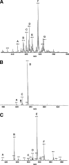
ESI-MS analysis of biliary bile salts from sea lamprey, zebrafish, and African clawed frog. From an extensive library generated from the analysis of many bile specimens, the major ions are annotated with probable matches indicating the number of carbon atoms (24 or 27), whether the compound is a bile acid or bile alcohol, the number of hydroxyl groups, the presence of a double bond (if any), and the type of conjugation. Additional studies (data not shown) included GC-MS, thin-layer chromatography, and high-performance liquid chromatography. The orientation of the hydrogen atom on carbon 5 was generally resolved by GC-MS. A: ESI-MS analysis of sea lamprey biliary bile. The labeled peaks in the spectra are as follows: peak A, C24 bile alcohol-(OH)2-SO4 with one double bond; peak B, C24 bile alcohol-(OH)3-SO4 with one double bond; peak C, C27 bile alcohol-(OH)2-SO4 with one double bond; peak D, C24 bile alcohol-(OH)4-SO4; peak E, C27 bile alcohol-(OH)3-SO4 with one double bond; peak F, C27 bile alcohol-(OH)4-SO4; peak G, C27 bile alcohol-(OH)5-SO4. Peak D corresponds to 5α-petromyzonol 24-sulfate. B: ESI-MS analysis of zebrafish biliary bile. The labeled peaks in the spectra are as follows: peak A, C24 bile acid-(OH)2-taurine; peak B, C24 bile acid-(OH)3-taurine; peak C, C27 bile alcohol-(OH)4-SO4; peak D, C27 bile alcohol-(OH)5-SO4. The most abundant ion (peak D) corresponds to 5α-cyprinol 27-sulfate. C: ESI:MS analysis of bile salts from African clawed frog biliary bile. The labeled peaks in the spectra are as follows: peak A, C24 bile acid-(OH)3; peak B, C27 bile acid-(OH)3; peak C, C24 bile acid-(OH)2-taurine; peak D, C24 bile acid-(OH)3-taurine; peak E, C27 bile alcohol-(OH)5-SO4; peak F, C27 bile acid-(OH)3-taurine. The most abundant ion (peak E) corresponds to 5β-cyprinol 27-sulfate, the A/B cis isomer to the main zebrafish bile salt.
Fig. 3.
Fig. 3.
Bile salt activation of farnesoid X receptors (FXRs) from different species. Frog and zebrafish FXRs are activated by the early bile salt 5α-cyprinol sulfate (A) but are insensitive to the recent primary bile salt 5β-chenodeoxycholyltaurine (TCDCA; B) and its corresponding deconjugated and dehydroxylated secondary bile salt 5β-lithocholic acid (C). In contrast, human and mouse FXRs are activated by 5β-bile acids (B, C) but not by 5α-cyprinol sulfate (A). The C. intestinalis FXR is insensitive to all three bile salts (A–C). Data shown are means ± SD.
Fig. 4.
Fig. 4.
Docking of ligands into structural models derived from X-ray crystallography or homology modeling. A: The curved binding pocket of human FXR accommodates the bent conformation of 5β-lithocholic acid well. B: The narrow binding pocket of sea lamprey FXR is suited to binding of the evolutionary early, planar bile salt 5α-cyprinol 27-sulfate. C: The binding pocket of Ciona FXR is smaller than that of any of the vertebrate FXRs analyzed and was not predicted to be capable of binding any bile salts but does bind the synthetic ligand AM-580. D: The large binding pocket of human pregnane X receptor (PXR) readily accommodates lithocholic acid. E: Similar to lamprey FXR, zebrafish PXR has a flat binding pocket suited to binding of the planar bile salt 5α-cyprinol 27-sulfate. F: Human vitamin D receptor has a relatively small, slightly curved binding pocket that can barely accommodate the small secondary bile acid lithocholic acid. See Experimental Procedures for details on the structural models.

References

    1. Hofmann A. F. 1999. The continuing importance of bile acids in liver and intestinal disease. Arch. Intern. Med. 159 2647–2658. - PubMed
    1. Gass J., H. Vora, A. F. Hofmann, G. M. Gray, and C. Khosla. 2007. Enhancement of dietary protein digestion by conjugated bile acids. Gastroenterology. 133 16–23. - PubMed
    1. Hofmann A. F., and L. Eckmann. 2006. How bile acids confer gut mucosal protection against bacteria. Proc. Natl. Acad. Sci. USA. 103 4333–4343. - PMC - PubMed
    1. Kalaany N. Y., and D. J. Mangelsdorf. 2006. LXRs and FXR: the yin and yang of cholesterol and fat metabolism. Annu. Rev. Physiol. 68 159–191. - PubMed
    1. Haslewood G. A. D. 1967. Bile salt evolution. J. Lipid Res. 8 535–550. - PubMed

Publication types