Review
. 2008 Apr;36(2):51-7.
doi: 10.1097/JES.0b013e318168e9dc.

Nuclear apoptosis contributes to sarcopenia

Affiliations
Review

Nuclear apoptosis contributes to sarcopenia

Stephen E Alway et al. Exerc Sport Sci Rev. 2008 Apr.

Abstract

Apoptosis results in DNA fragmentation and, subsequently, destruction of cells containing a single nucleus. Our hypothesis is that multinucleated cells such as muscle fibers can experience apoptotic-induced loss of single nuclei (nuclear apoptosis) without destruction of the entire fiber. The loss of nuclei likely contributes to atrophy and sarcopenia. Furthermore, increased chronic activity attenuates apoptotic signaling, which may reduce sarcopenia.


Figures
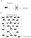
Figure 1
Figure 1
Apoptosis and nuclear apoptosis. A. Apoptosis results in nuclear fragmentation and elimination of the nuclei and death of single cells. B. Our hypothesis is that nuclear apoptotic signaling targets specific myonuclei in a muscle fiber. Elimination of myonuclei results in a decrease in the number of nuclei in the muscle fiber and, therefore, an increase in the cytoplasmic volume per nucleus. The muscle fiber undergoes atrophy to accommodate the fewer available nuclei for transcriptional control in an attempt to reestablish the original cytoplasmic volume per nucleus.
Figure 2
Figure 2
Involvement of nuclear apoptosis in sarcopenia during loading and unloading (A–C). Our hypothesis is that apoptotic signaling targets specific MN in multinucleated muscle fiber cells (nuclei targeted for apoptosis are indicated by an “X”) without inflammation or disturbing adjacent proteins or organelles in aging (A). Elimination of nuclei results in a decrease in the total number of nuclei available to sustain the cytoplasmic volume, and this increases the cytoplasmic volume per nucleus (B). The muscle fiber undergoes atrophy to accommodate the fewer available nuclei for transcriptional control. This is an attempt to reestablish the original cytoplasmic volume per nucleus (C). If a sufficient number of nuclei are targeted for death in the multinucleated muscle fiber, the entire cell will be eliminated (D–F). Sarcopenia can be partially offset by exercise and muscle loading (e.g., by resistance exercise), such that loading of the original muscle fibers results in a stimulus that (D) results in a proliferation of SC, which increases the number of nuclei and reduces the fiber cytoplasm volume per nucleus (E). However, proliferation of satellite cells is largely attenuated in aging compared with young muscles. The decreased cytoplasmic volume–to–nucleus ratio provides a stimulus to induce fiber hypertrophy in an attempt to reestablish the original cytoplasmic volume per nucleus ratio (F). As aged muscle fibers have fewer total nuclei than young muscles, aged muscle fibers cannot hypertrophy to the same absolute size as in young muscle (G–J). If the hypertrophied muscle is now unloaded (e.g., stop exercise training, prolonged bed rest, etc.), nuclei are targeted for apoptosis, but nuclear apoptosis occurs preferentially (at least initially) in the most recently activated satellite cells (G). This reduces the total number of nuclei and increases cytoplasmic volume per nucleus ratio (H). Fibers atrophy to reestablish the original cytoplasmic volume per nucleus ratio, but fiber atrophy is more pronounced in aging (I). Nuclear apoptosis continues to target nuclei (J) as a result of an aging-induced apoptotic signaling environment, and this further exacerbates muscle fiber atrophy (C), which compounds muscle loss associated with sarcopenia. MN indicates myonuclei; SC, satellite cells.
Figure 3
Figure 3
Apoptotic signaling in aging muscles. This figure summarizes the extrinsic death domain pathway and intrinsic pathway for mitochondrial apoptosis in aging muscles under conditions of muscle loss (e.g., unloading, denervation). The increase (+) and the decrease (−) in the direction of the change in apoptotic signaling in response to muscle wasting insults for proteins in these pathways are indicated in muscles from young adult (black) and old (gray) animals. Double positive (++) or double negative (−−) signs indicate a large increase or decrease in protein levels, respectively. Proapoptotic pathways are shown by lines and arrows. A line with a blunt end indicates an antiapoptotic influence. Dotted lines show examples of proapoptotic proteins that are released from the mitochondria in response to muscle wasting, which can either directly (e.g., AIF) or indirectly (e.g., cytochrome c) induce nuclear apoptosis in muscle cells. TRADD, tumor necrosis factor receptor 1 associated death domain.

References

    1. Allen DL, Roy RR, Edgerton VR. Myonuclear domains in muscle adaptation and disease. Muscle Nerve. 1999;22:1350–1360. - PubMed
    1. Alway SE, Degens H, Krishnamurthy G, Smith CA. Potential role for Id myogenic repressors in apoptosis and attenuation of hypertrophy in muscles of aged rats. Am. J. Physiol. Cell Physiol. 2002;283:C66–C76. - PubMed
    1. Alway SE, Siu PM, Murlasits Z, Butler DC. Muscle hypertrophy models: applications for research on aging. Can. J. Appl. Physiol. 2005;30:591–624. - PubMed
    1. Brack AS, Bildsoe H, Hughes SM. Evidence that satellite cell decrement contributes to preferential decline in nuclear number from large fibres during murine age-related muscle atrophy. J. Cell Sci. 2005;118(pt 20):4813–4821. - PubMed
    1. Bruusgaard JC, Liestol K, Gundersen K. Distribution of myonuclei and microtubules in live muscle fibers of young, middle-aged, and old mice. J. Appl. Physiol. 2006;100:2024–2030. - PubMed

Publication types