. 2008 Apr;40(4):460-5.
doi: 10.1038/ng.112. Epub 2008 Mar 23.

Newly identified prion linked to the chromatin-remodeling factor Swi1 in Saccharomyces cerevisiae

Affiliations

Newly identified prion linked to the chromatin-remodeling factor Swi1 in Saccharomyces cerevisiae

Zhiqiang Du et al. Nat Genet. 2008 Apr.

Abstract

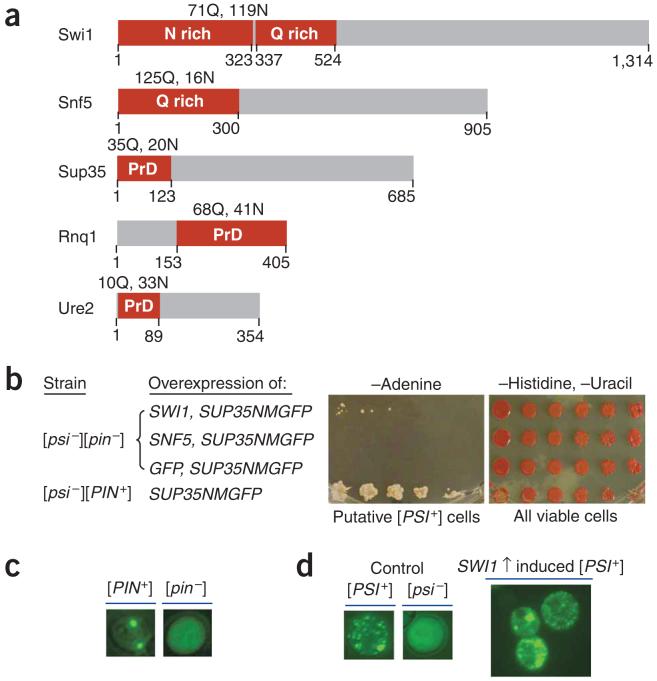
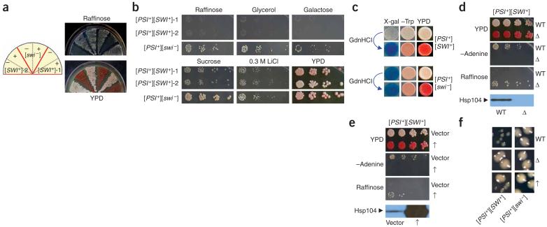
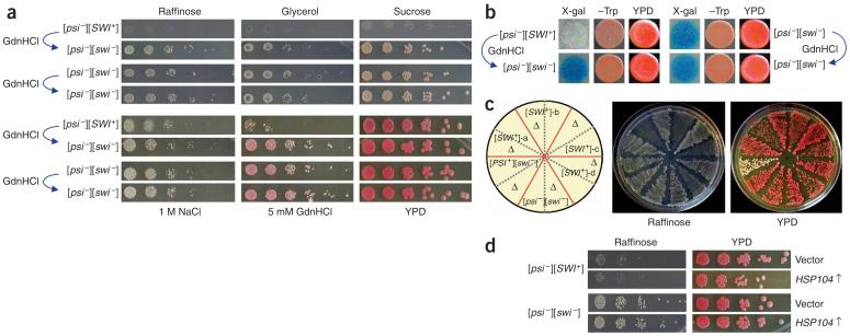
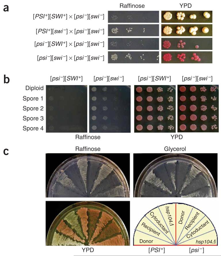
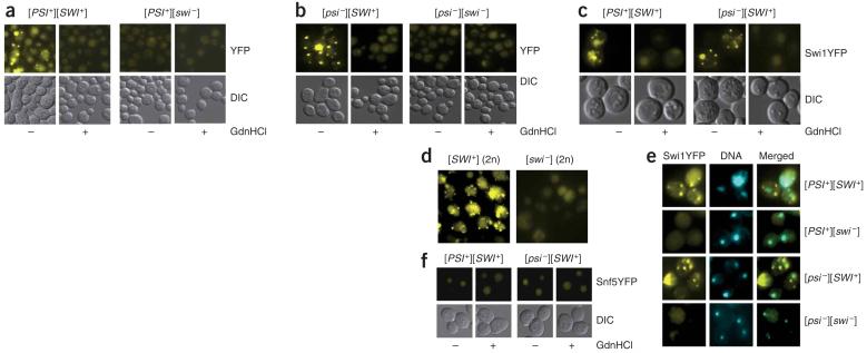
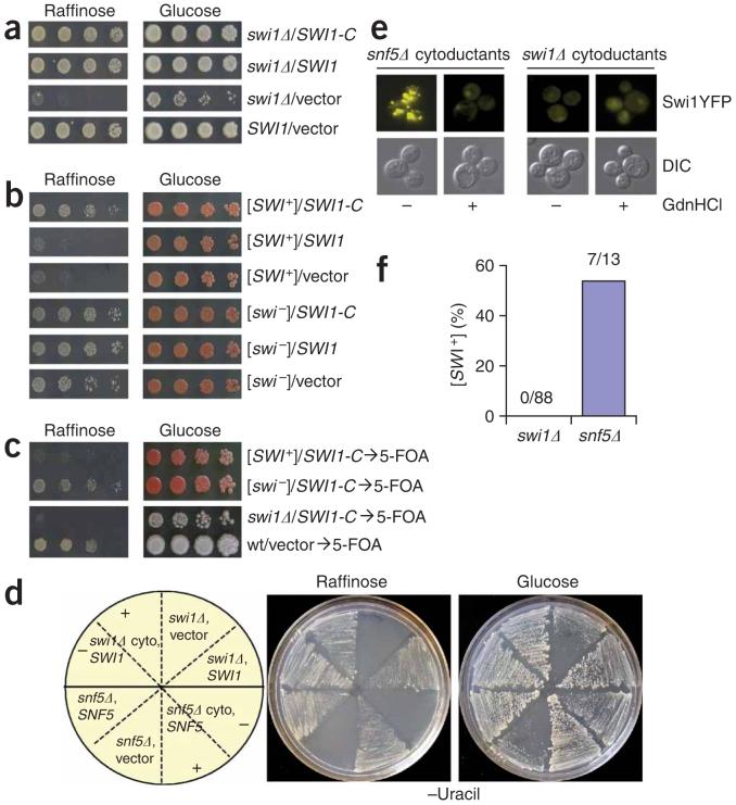
SWI/SNF, an evolutionarily conserved ATP-dependent chromatin-remodeling complex, has an important role in transcriptional regulation. In Saccharomyces cerevisiae, SWI/SNF regulates the expression of approximately 6% of total genes through activation or repression. Swi1, a subunit of SWI/SNF, contains an N-terminal region rich in glutamine and asparagine, a notable feature shared by all characterized yeast prions--a group of unique proteins capable of self-perpetuating changes in conformation and function. Here we provide evidence that Swi1 can become a prion, [SWI+]. Swi1 aggregates in [SWI+] cells but not in nonprion cells. Cells bearing [SWI+] show a partial loss-of-function phenotype of SWI/SNF. [SW+] can be eliminated by guanidine hydrochloride treatment, HSP104 deletion or loss of Swi1. Moreover, we show [SWI+] is dominantly and cytoplasmically transmitted. Our findings reveal a novel mechanism of 'protein-only' inheritance that results in modification of chromatin-remodeling and, ultimately, global gene regulation.


Figures

Figure 1
Figure 1
Overexpression of SWI1 but not SNF5 functions as Pin+. (a) Diagrams of Swi1 and Snf5 with their glutamine (Q) and asparagine (N) rich regions shown (red boxes). The prion domains (PrDs) of Sup35, Rnq1 and Ure2 are also indicated (red boxes). (b) Overexpression of SWI1 can substitute for [PIN+] to promote [PSI+] formation. We grew 74D-694 cells [(psi-][pin-]) containing pRS313CUP1NMGFP and p426GPDSWI1, pRS313CUP1NMGFP and p426GPDSNF5, or pRS313CUP1NMGFP and p426GPDGFP to early-log phase and induced them for NMGFP expression by addition of CuSO4 to 100 μM. After 24-h induction, cells were spotted onto the indicated plates with a fivefold serial dilution. Pictures were taken after 5-d incubation for cells on synthetic complete medium lacking histidine and uracil (-Histidine, -Uracil) or after 9 d for cells on medium lacking adenine (-Adenine). (c) Fluorescence microscopy assay of Rnq1 conformational status. Before conducting experiments shown in panel b, we confirmed the Rnq1 conformational status by transforming with p416CUP1RNQ1GFP, inducing for 4 h with 100 μM CuSO4 and analyzing by fluorescence microscopy. (d) Sup35 is aggregated in Ade+ cells obtained by SWI1 and SUP35NMGFP co-overexpression. After eliminating p426GPDSWI1 and pRS313CUP1NMGFP, we transformed Ade+ isolates from panel b with p416CUP1NMGFP and analyzed them by fluorescence microscopy to confirm their [PSI+] status. Shown here are fluorescence patterns of a representative [PSI+] isolate and isogenic [PSI+] and [psi-] controls.
Figure 2
Figure 2
Isolation and characterization of [SWI+] candidates. (a) [SWI+]-1 and [SWI+]-2, two [SWI+] candidates isolated from [PSI+] cells upon co-overexpression of SUP35NMGFP and SWI1, showed a guanidine hydrochloride-curable Raf- phenotype. Shown are cell streaks of the candidates before (-) and after (+) guanidine hydrochloride (GdnHCl) treatment on indicated media. Note that both of the plasmids, pRS313CUP1NMGFP and p426GPD-SWI1, were eliminated before 5 mM guanidine hydrochloride treatment. (b) Phenotypic assays of the [PSI+][SWI+] candidates. [SWI+] candidates and [SWI-] control were grown in YPD to mid-log phase and then spotted to the indicated media with a fivefold serial dilution. (c) [SWI+]-2 (upper) and [SWI-] (lower) cells containing pLS7 plasmid (Trp+), a SWI/SNF lacZ reporter controlled by a chimeric promoter composed of the SWI/SNF regulatory sequence of SUC2 promoter and the core promoter of LEU2 (ref. 24), were spotted onto +sucrose -tryptophan synthetic complete media + 20 μg/ml X-gal plates (X-gal); +glucose -tryptophan synthetic complete medium plates (-Trp); and YPD (YPD) plates. (d) HSP104 deletion eliminated [PSI+] and the Raf- phenotype. [SWI+]-2 cells with (Δ) or without (WT) HSP104 disruption were assayed on indicated media. (e) The effect of HSP104 overexpression on Raf- phenotype. [SWI+]-2 cells containing p2HGHSP104 (↑) or p2HG (vector) were grown in synthetic complete liquid media lacking histidine and then spotted to the indicated media. HSP104 disruption and overexpression were confirmed by immunoblot analysis using a polyclonal Hsp104 antibody. (f) Enlarged images showing the colony sizes of [PSI+][SWI+] and [PSI+][SWI-] cells after HSP104 deletion (Δ), overexpression (↑) or no treatment (WT). A [PSI+] isolate with Raf+ phenotype obtained under identical conditions for [SWI+] candidate isolation was used as the nonprion ([SWI-]) control in all experiments described in this figure.
Figure 3
Figure 3
[SWI+] can exist independently from [PSI+]. (a) Phenotypic analysis of a representative [psi-][SWI+] isolate derived from [PSI+][SWI+]-2 upon Hsp104 overproduction. After eliminating the HSP104 overexpression plasmid, [psi-][SWI+] cells treated with or without 5 mM guanidine hydrochloride were spotted onto indicated plates with a fivefold serial dilution. Pictures were taken after 5-d incubation at 30 °C. (b) Isogenic [psi-][SWI+] and [psi-][SWI-] cells containing pLS7 were treated with or without 5 mM guanidine hydrochloride and were grown in -tryptophan synthetic complete liquid media to mid-log phase before spotting to +sucrose -tryptophan media + 20 μg/ml X-gal (X-gal), +glucose -tryptophan media (-Trp) and YPD (YPD) plates. (c) HSP104 disruption eliminates [SWI+] in [psi-][SWI+] isolates. Shown are cell streaks of four isolates on YPD and raffinose plates before and after HSP104 gene disruption. Δ, HSP104 disruption. (d) The [SWI+] prion is tolerant to Hsp104 overproduction. Isogenic [psi-][SWI+] and [psi-][SWI-] cells containing p2HG (vector) or p2HGHSP104 (HSP104↑) were cultured and spotted to the indicated plates.
Figure 4
Figure 4
[SWI+] is dominantly and cytoplasmically inherited. (a) [SWI+] diploids show Raf- phenotype. Diploid cells derived from the indicated crosses were grown in YPD to mid-log phase and spotted onto a raffinose or YPD plate with a fivefold serial dilution. (b) All meiotic progenies of [SWI+] show the Raf- phenotype. Shown are representative phenotypic assays of a full set of spores derived from isogenic [SWI+] or [SWI-] diploids. (c) [SWI+] cytoductants of c10B-H49 showed the Raf- phenotype that could be eliminated by hsp104Δ. Shown are donors, 74D-694 [PSI+][SWI+] (left) or [psi-][SWI+] (right); recipient, c10B-H49 [psi-][SWI-]; cytoductants, [PSI+][SWI+] (left) or [psi-][SWI+] (right); and the hsp104Δ derivatives of the corresponding cytoductants.
Figure 5
Figure 5
Swi1 but not Snf5 exists in distinct conformational states in [SWI+] and [swi-] cells. (a,b) Swi1-YFP was aggregated in [PSI+][SWI+] (a) and [psi-][SWI+] (b) but not in isogenic [swi-] cells or [SWI+] cells after guanidine hydrochloride treatment. Cells as indicated were treated with (+) or without (-) 5 mM guanidine hydrochloride. (c) c10B-H49 cytoductants of [SWI+] showed guanidine hydrochloride-curable fluorescent foci of Swi1-YFP. (d) Swi1-YFP was aggregated in the 74D-694 [psi-][SWI+] diploids but not in [psi-][swi-] diploids. (e) Swi1-YFP aggregates were mainly localized in cytoplasm. We stained 74D-694 [SWI+] cells in both [PSI+] and [psi-] backgrounds with Hoechst dye and analyzed them as described in Methods. (f) Endogenous Snf5-YFP is not aggregated in [SWI+] or [swi-] cells. Cells were treated with (+) or without (-) 5 mM guanidine hydrochloride and analyzed under a fluorescence microscope. All cells were transformed with p416TEFSWI1YFP and analyzed by fluorescence microscopy assays after growth in synthetic complete medium lacking uracil to mid-log phase.
Figure 6
Figure 6
Swi1 is the protein determinant of [SWI+]. (a) The C domain is sufficient for Swi1 function. Shown are S288C swi1Δ cells containing p416TEFSWI1-C (SWI1-C), p416TEFSWI1 (SWI1) or p416TEF (vector). Wild-type S288C with p416TEF was included as a control. (b) SWI1-C, but not the full-length SWI1, was able to ‘complement’ the Raf- phenotype of [SWI+]. Cells of 74D-694 [PSI+][SWI+] and [PSI+][swi-] containing p416TEFSWI-C (SWI1-C), p416TEFSWI1 (SWI1) or p416TEF (vector) were examined for growth on the indicated plates. (c) Swi1-C was able to phenotypically ‘mask’ but not cure [SWI+]. After plasmid removal, [SWI+] and swi1Δ cells were re-assayed as described in a and b. (d) Swi1 is required for [SWI+] propagation. [SWI+] was cytoduced to isogenic swi1Δ and snf5Δ cells, and the resulting cytoductants were transformed with a SWI1 expression plasmid for swi1Δ cells (swi1Δ cyto, SWI1) or SNF5 for snf5Δ cells (snf5Δ cyto, SNF5). Isogenic swi1Δ and snf5Δ cells were also included as controls. Cell streaks of each representative transformant before (-) or after (+) 5 mM guanidine hydrochloride treatment are shown on the indicated plates. (e) Swi1-YFP is aggregated in the snf5Δ but not in the swi1Δ cytoductants after [SWI+] cytoduction. Cytoductants of swi1Δ and snf5Δ were treated with (+) or without (-) 5 mM guanidine hydrochloride followed by transformation of p416TEF-SWI1YFP and analysis by fluorescence microscopy. (f) Summary of the [SWI+] cytoduction experiments. Shown are the results of three independent experiments.

References

    1. Martens JA, Winston F. Recent advances in understanding chromatin remodeling by Swi/Snf complexes. Curr. Opin. Genet. Dev. 2003;13:136–142. - PubMed
    1. Sudarsanam P, Iyer VR, Brown PO, Winston F. Whole-genome expression analysis of snf/swi mutants of Saccharomyces cerevisiae. Proc. Natl. Acad. Sci. USA. 2000;97:3364–3369. - PMC - PubMed
    1. Tuite MF, Cox BS. Propagation of yeast prions. Nat. Rev. Mol. Cell Biol. 2003;4:878–890. - PubMed
    1. Wickner RB. [URE3] as an altered Ure2 protein: evidence for a prion analog in Saccharomyces cerevisiae. Science. 1994;264:566–569. - PubMed
    1. Prusiner SB. Prions. Proc. Natl. Acad. Sci. USA. 1998;95:13363–13383. - PMC - PubMed

Publication types

MeSH terms