. 2008 Jun;88(6):602-14.
doi: 10.1038/labinvest.2008.25. Epub 2008 Mar 24.

Motility-related actinin alpha-4 is associated with advanced and metastatic ovarian carcinoma

Affiliations

Motility-related actinin alpha-4 is associated with advanced and metastatic ovarian carcinoma

Maria V Barbolina et al. Lab Invest. 2008 Jun.

Abstract

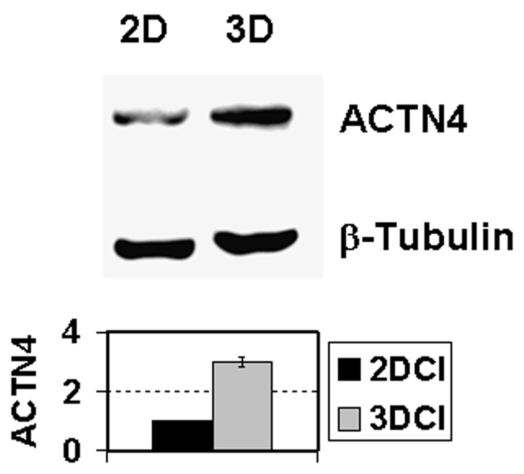
Advanced and metastatic ovarian cancer is a leading cause of death from gynecologic malignancies. A more detailed understanding of the factors controlling invasion and metastasis may lead to novel anti-metastatic therapies. To model cellular interactions that occur during intraperitoneal metastasis, comparative cDNA microarray analysis and confirmatory real-time reverse transcription PCR (RT-PCR) were employed to uncover changes in gene expression that may occur in late stage ovarian cancer in response to microenvironmental cues, particularly native three-dimensional collagen I. Gene expression in human ovarian carcinoma tissues was evaluated on the RNA and protein level using real-time RT-PCR and immunohistochemistry. Cell invasion and migration were evaluated in a collagen invasion assay and a scratch wound assay. Three-dimensional collagen I culture led to differential expression of several genes. The role of actinin alpha-4 (ACTN4), a cytoskeleton-associated protein implicated in the regulation of cell motility, was examined in detail. ACTN4 RNA and protein expression were associated with advanced and metastatic human ovarian carcinoma. This report demonstrates that a cytoskeletal-associated protein ACTN4 is upregulated by three-dimensional collagen culture conditions, leading to increased invasion and motility of ovarian cancer cells. Expression of ACTN4 in human ovarian tumors was found to be associated with advanced-stage disease and peritoneal metastases.


Figures

Figure 1
Figure 1. 3-dimensional collagen culture induces changes in gene expression
Real time RT-PCR analysis of expression of genes (A) upregulated and (B) downregulated by the three-dimensional collagen I culture compared to two-dimensional collagen I. Actinin alpha-4 (ACTN4), jun B proto-oncogene (JUNB), MAX interactor 1 (MXI1), solute carrier family 20 (SLC20A1), tumor necrosis factor (ligand) superfamily member 10 (TNFSF10), claudin-1 (CLDN1), hematopoietically expressed homeobox (HHEX), interleukin 6 (IL6), and transforming growth factor beta 2 (TGFB2) expression was examined in cells cultured cultured on thin-layer (2D) and three-dimensional (3D) collagen I gel for 8 hours. Total RNA was purified, cDNA synthesized, and quantitative real time RT-PCR carried out as described in Methods. Results are plotted as the expression ratio, indicating the fold change of mRNA expression in cells cultured on (A) 3DCI relative to thin layer collagen or (B) thin layer collagen relative to 3DCI. Data are shown as mean ± standard deviation, and are an average of three independent experiments.
Figure 2
Figure 2. Actinin alpha-4 protein is upregulated by three-dimensional collagen culture
Cells were cultured on 3DCI and 2DCI for 24 hours followed by Western blot analysis of the cell lysates for ACTN4 expression. β-Tubulin was used as a loading control. Primary antibodies against ACTN4 and β-tubulin were used at 1:200 and 1:2000 dilutions, respectively. Secondary peroxidase conjugated anti-goat and anti-mouse IgG were used at 1:1000 and 1:2000 dilutions to visualize ACTN4 and β-tubulin. The histogram shows the levels of ACTN4 expression in DOV13 cultured on 2DCI and 3DCI relative to the β-tubulin expression. The data represent an average of three independent experiments with a standard deviation ± 5%.
Figure 3
Figure 3. Real time RT-PCR analysis of ACTN4 mRNA expression in normal ovarian epithelium and epithelial ovarian carcinomas of various histological type and stage of tumorigenic development
(A) ACTN4 expression in ovarian carcinoma tissues and normal ovarian epithelia. (B, C) Expression of two housekeeping genes, β-actin (ACTNB) and ribosomal protein L19 (RPL-19), respectively. Black bars in the histograms represent the values of the cycle numbers at which the accumulation of fluorescent signal from SYBR Green bound to the gene-specific double-stranded DNA PCR product was above the background. Absence of the black bars indicates no accumulation of the PCR product specific to ACTN4. A total of 48 samples was tested for the expression of ACTN4, ACTNB, and RPL-19. Results for samples from 1–48 are plotted from left to right on the x axis and separated by small ticks. Samples from normal epithelia are designated ‘0’, and those belonging to ovarian carcinoma FIGO stages I, II, III, IV, are indicated as ‘I’, ‘II’, ‘III’, ‘IV’, respectively, and are separated by long ticks.
Figure 4
Figure 4. Immunohistochemical analysis of ACTN4 expression in paired primary and metastatic human epithelial ovarian carcinomas
(A,B) Representative example of paired (A) primary tumor and (B) the corresponding metastatic lesion from the same patient. Tissues were stained with ACTN4 specific antibodies as indicated in Methods. In both primary and metastatic specimens, cytoplasmic staining with ACTN4 antibodies was detected (ACTN4 – brown; nuclear DNA – hematoxylin&eosin); Inserts show magnified representative portions of the stained tissue.
Figure 5
Figure 5. ACTN4 gene silencing retards ovarian cancer invasion through three-dimensional collagen
(A) Boyden chamber 3D collagen invasion assay. DOV13 cells were transfected with siRNA for ACTN4 or control siRNA, as indicated, and invasion of collagen gels was evaluated in a modified Boyden chamber assay as described in Methods. Histograms show a percentage of invading cells, with invasion of wild type cells designated as 100%. Data represent an average of 3 independent experiments. Representative bright field images of cells invading to the underside of the filter are shown below the histograms. (B) ACTN4 does not affect MT1-MMP expression (upper panel). Cells were transfected with ACTN4 and control siRNA, and the cell lysates were examined for MT1-MMP expression by Western blot (1:1000 dilution of anti-MT1-MMP, 1:4000 dilution of peroxidase conjugated anti-rabbit IgG). ACTN4 siRNA is efficient in silencing the ACTN4 gene expression (middle panel) (1:200 dilution of anti-ACTN4, 1:1000 dilution of peroxidase conjugated anti-goat IgG). The histogram at the bottom demonstrates levels of ACTN4 expression in untransfected DOV13 (black bar) compared to those transfected with control siRNA (dark grey bar) and ACTN4 siRNA (light grey bar). The data represent an average of two independent experiments with a standard deviation ± 10%. The asterisk shows significant differences to the control with p≤0.005.
Figure 6
Figure 6. ACTN4 gene silencing retards ovarian cancer cell motility
(A) Scratch wound motility assay. DOV13 cell monolayers were wounded and observed over the indicated periods of time, 25 and 50 hours. Note that cell motility of DOV13 transfected with phosphate buffered saline was the same as that for cells transfected with the control siRNA, and, hence, the former are not shown in the picture. Photographs of the wounds are representative of three independent experiments that were performed and quantified as indicated in Methods and are taken at 5x magnification. (B) Histograms illustrate the percentage of cell motility in the wound healing assay with the standard deviation of ±10%. Dark grey and light grey bars represent % of wound healing in cell monolayers transfected with control and ACTN4 siRNA, respectively. * p≤0.006, ** p≤0.012. (C) ACTN4 is upregulated in cells on the edge of the healing wound. Wounds were introduced in cell monolayers cultured in standard conditions on glass coverslips coated with collagen I, allowed to migrate for 20 h, fixed and stained with DAPI and ACTN4-specific antibody as indicated in the Methods. Photographs of cells in phase, DAPI- and ACTN4-fluorescence, were taken at 5× magnification on the objective using a Zeiss Axiovert fluorescent microscope, as indicated. White arrows indicate the position of the migrating edge of the cell monolayer and an increase in ACTN4 expression in migrating cells. (D) Cytoplasmic staining of ACTN4 in migrating cells. Cells cultured on collagen-coated glass coverslips were induced to migrate in a scratch wound assay for 20 h, fixed and stained with ACTN4 antisera (1:100 dilution), Alexa 488 phalloïdin (red), and DAPI (blue) as indicated in Methods. Photographs were taken with Zeiss Axiovert fluorescent microscope at 40× magnification on the objective. White arrows show cytoplasmic localization of ACTN4 in migrating cells. A white line is drawn starting from the migrating edge (marked with a cyan dot) toward the monolayer (marked with a yellow dot) to compare the intensity of intracellular ACTN4 staining. Intensity of the ACTN4 staining is quantified using ImageJ (NIH). Cyan and yellow dots on the graph correspond to the beginning and the end of the line drawn to generate the intensity scan.

References

    1. Dupont NC, Berman ML. Surgical management of epithelial ovarian cancer: a review of the literature. Minerva Ginecologica. 2004;56(6):547–556. - PubMed
    1. Guppy AE, Nathan PD, Rustin GJ. Epithelial ovarian cancer: a review of current management. Clinical Oncology (Royal College of Radiologists) 2005;17(6):399–411. - PubMed
    1. Jemal A, Siegel R, Ward E, Murray T, Xu J, Smigal C, et al. Cancer statistics, 2006. CA: a Cancer Journal for Clinicians. 2006;56(2):106–130. - PubMed
    1. Runnebaum IB, Stickeler E. Epidemiological and molecular aspects of ovarian cancer risk. Journal of Cancer Research & Clinical Oncology. 2001;127(2):73–79. - PubMed
    1. Sankaranarayanan R, Ferlay J. Worldwide burden of gynaecological cancer: the size of the problem. Best Practice & Research in Clinical Obstetrics & Gynaecology. 2006;20(2):207–225. - PubMed

Publication types

MeSH terms