Review
. 2008 Apr;36(Pt 2):141-7.
doi: 10.1042/BST0360141.

Biochemical and structural analysis of alpha-catenin in cell-cell contacts

Affiliations
Review

Biochemical and structural analysis of alpha-catenin in cell-cell contacts

Sabine Pokutta et al. Biochem Soc Trans. 2008 Apr.

Abstract

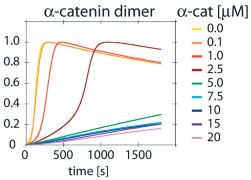
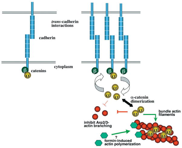
Cadherins are transmembrane adhesion molecules that mediate homotypic cell-cell contact. In adherens junctions, the cytoplasmic domain of cadherins is functionally linked to the actin cytoskeleton through a series of proteins known as catenins. E-cadherin binds to beta-catenin, which in turn binds to alpha-catenin to form a ternary complex. alpha-Catenin also binds to actin, and it was assumed previously that alpha-catenin links the cadherin-catenin complex to actin. However, biochemical, structural and live-cell imaging studies of the cadherin-catenin complex and its interaction with actin show that binding of beta-catenin to alpha-catenin prevents the latter from binding to actin. Biochemical and structural data indicate that alpha-catenin acts as an allosteric protein whose conformation and activity changes depending on whether or not it is bound to beta-catenin. Initial contacts between cells occur on dynamic lamellipodia formed by polymerization of branched actin networks, a process controlled by the Arp2/3 (actin-related protein 2/3) complex. alpha-Catenin can suppress the activity of Arp2/3 by competing for actin filaments. These findings lead to a model for adherens junction formation in which clustering of the cadherin-beta-catenin complex recruits high levels of alpha-catenin that can suppress the Arp2/3 complex, leading to cessation of lamellipodial movement and formation of a stable contact. Thus alpha-catenin appears to play a central role in cell-cell contact formation.


Figures

Figure 1
Figure 1. Standard model of junction organization
The cytoplasmic domain of classical cadherins binds to β-catenin. β-Catenin in turn binds to α-catenin, which binds to F-actin either directly or through other actin-binding proteins.
Figure 2
Figure 2. Primary structure of α-catenin, with vinculin-homology regions and binding sites for various partners indicated
Numbers are amino acid positions. ZO-1, zonula occludens 1.
Figure 3
Figure 3. α-Catenin homodimer structure and binding to β-catenin
(A) Crystal structure of the homodimerization domain, with protomers coloured red and turquoise. (B) Schematic illustration of β-catenin binding to α-catenin 57–264. The homodimerization domain 82–264 is shown in yellow, and the flexibly linked α-catenin 57–81 sequence in blue. The α-catenin-binding sequence of β-catenin, residues 118–149, forms a helix and is shown in red. (C) Diagram of the βα-catenin chimaera used to form a stable β-catenin–α-catenin heterodimer, coloured as in (B). (D) Crystal structure of the βα-catenin chimaera, coloured as in (B). Adapted from Mol. Cell, 5, Pokutta, S. and Weis, W.I., ‘Structure of the dimerization and β-catenin binding region of α-catenin’, 533–543, © 2000, with permission from Elsevier.
Figure 4
Figure 4. Summary of FRAP parameters for E-cadherin, β-catenin, α-catenin and actin
Ecad, E-cadherin–GFP; EcadΔC, E-cadherin–GFP with the cytoplasmic domain deleted; αcat, α-catenin–GFP; αcatΔC, α-catenin–GFP with the actin-binding domain deleted; βcat, β-catenin–GFP; Actin, actin–GFP; Rh-actin, rhodamine-labelled actin microinjected into cells. Results are means ± S.E.M. Adapted from Cell, 123, Yamada, S., Pokutta, S., Drees, F., Weis, W.I. and Nelson, W.J. ‘Deconstructing the cadherin–catenin–actin complex’, 889–901, © 2005, with permission from Elsevier.
Figure 5
Figure 5. α-Catenin can suppress Arp2/3-mediated actin polymerization
Increasing amounts of α-catenin were added to a solution containing pyrene-labelled actin and 50 nM Arp2/3 and the stimulatory WASP (Wiskott–Aldrich syndrome protein) VCA (verprolin homology, cofilin homology and acidic) domain. Polymerization results in an increase in fluorescence. Adapted from Cell, 123, Drees, F., Pokutta, S., Yamada, S., Nelson, W.J. and Weis, W.I. ‘α-Catenin is a molecular switch that binds E-cadherin/β-catenin and regulates actin filament assembly’, 903–915, © 2005, with permission from Elsevier.
Figure 6
Figure 6. Model for cell–cell contact formation
Transient contacts of cadherins on lamellipodia (upper left) lead to clustering of the cadherin–β-catenin complex (upper right) that recruits α-catenin. Exchange of α-catenin between cadherin-bound β-catenin and cytosol results in high local concentration of α-catenin, favouring dimerization (right). Dimeric α-catenin near the membrane can locally suppress Arp2/3 and shut off lamellipodial movement, generating a stable contact. The α-catenin may also help to cross-link actin and promote linear cable formation through formins.

References

    1. Farquhar MG, Palade GE. Junctional complexes in various epithelia. J Cell Biol. 1963;17:375–412. - PMC - PubMed
    1. Hirano S, Nose A, Hatta K, Kawakami A, Takeichi M. Calcium-dependent cell–cell adhesion molecules (cadherins): subclass specificities and possible involvement of actin bundles. J Cell Biol. 1987;105:2501–2510. - PMC - PubMed
    1. Lubarsky B, Krasnow MA. Making and shaping biological tubes. Cell. 2002;112:19–28. - PubMed
    1. Ehrlich JS, Hansen MD, Nelson WJ. Spatio-temporal regulation of Rac1 localization and lamellipodia dynamics during epithelial cell–cell adhesion. Dev Cell. 2002;3:259–270. - PMC - PubMed
    1. Chu YS, Thomas WA, Eder O, Pincet F, Perez E, Thiery JP, Dufour S. Force measurements in E-cadherin-mediated cell doublets reveal rapid adhesion strengthened by actin cytoskeleton remodeling through Rac and Cdc42. J Cell Biol. 2004;167:1183–1194. - PMC - PubMed

Publication types