. 2008 Dec;12(6B):2609-21.
doi: 10.1111/j.1582-4934.2008.00312.x. Epub 2008 Mar 19.

The induction of bone formation by smart biphasic hydroxyapatite tricalcium phosphate biomimetic matrices in the non-human primate Papio ursinus

Affiliations

The induction of bone formation by smart biphasic hydroxyapatite tricalcium phosphate biomimetic matrices in the non-human primate Papio ursinus

U Ripamonti et al. J Cell Mol Med. 2008 Dec.

Abstract

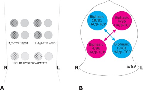
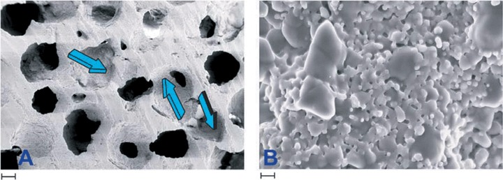
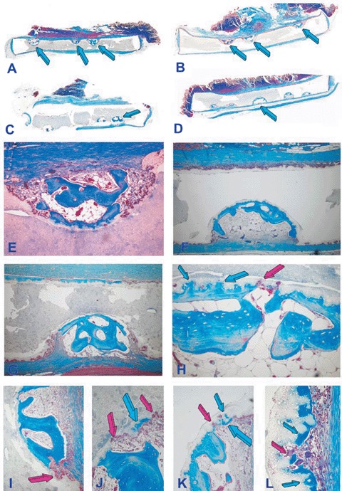
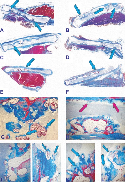
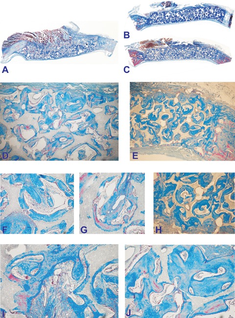
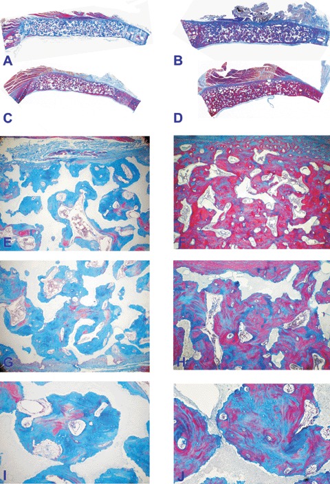
Long-term studies in the non-human primate Chacma baboon Papio ursinus were set to investigate the induction of bone formation by biphasic hydroxyapatite/p-tricalcium phosphate (HA/beta-TCP) biomimetic matrices. HA/beta-TCP biomimetic matrices in a pre-sinter ratio (wt%) of 40/60 and 20/80, respectively, were sintered and implanted in the rectus abdominis and in calvarial defects of four adult baboons. The post-sinter phase content ratios were 19/81 and 4/96, respectively. Morphological analyses on day 90 and 365 showed significant induction of bone formation within concavities of the biomimetic matrices with substantial bone formation by induction and resorption/dissolution of the implanted matrices. One year after implantation in calvarial defects, 4/96 biphasic biomimetic constructs showed prominent induction of bone formation with significant dissolution of the implanted scaffolds. The implanted smart biomimetic matrices induce de novo bone formation even in the absence of exogenously applied osteogenic proteins of the transforming growth factor-beta(TGF-beta) superfamily. The induction of bone formation biomimetizes the remodelling cycle of the cortico-cancellous bone of primates whereby resorption lacunae, pits and concavities cut by osteoclastogenesis are regulators of bone formation by induction. The concavities assembled in HA/beta-TCP biomimetic bioceramics are endowed with multifunctional pleiotropic self-assembly capacities initiating and promoting angiogenesis and bone formation by induction. Resident mesenchymal cells differentiate into osteoblastic cell lines expressing, secreting and embedding osteogenic soluble molecular signals of the TGF-beta superfamily within the concavities of the biomimetic matrices initiating bone formation as a secondary response.


Figures

Fig. 1
Fig. 1
Heterotopic rectus abdominisand orthotopic calvarial models in 4 adult Chacma baboons Papio ursinusfor bone induction and morphogenesis by sintered biphasic hydroxyapatite (HA) tricalcium phosphate (TCP) with post-sinter content ratios of 19/81 and 4/96, respectively, implanted in both heterotopic rectus abdominisand orthotopic calvarial sites. (A) Heterotopic intramuscular model and implantation design in the rectus abdominis muscle. HA/TCP 19/81, HA/TCP 4/96 and solid hydroxyapatite discs of 20 mm in diameter, 3 mm thickness with 25 hemispherical indentations on one planar surface only were implanted in quadruplicate two samples with the concavities facing ventrally and two samples with concavities facing dorsally to evaluate the extent of bone induction as directed by the site of implantation within the rectus abdominismuscle. (B) The calvarial Latin block design resulted in the contra lateral implantation of macroporous discs of either post-sinter 19/81 HA/TCP and 4/96 HA/TCP biphasic biomimetic matrices, two macroporous implants per animal for a total of 8 19/81 and 8 4/96 HA/TCP biphasic macroporous discs.
Fig. 2
Fig. 2
Scanning electron microphotographs of a specimen of hydroxyapatite/tricalcium phosphate 19/81. (A) Blue arrows show repetitive sequences of concavities with defined radii of curvature and diameters in macroporous specimens for orthotopic calvarial implantation. (B) Microstructure of the 19/81 biomimetic biphasic constructs. Larger grains are β-tricalcium phosphate, the smaller grains hydroxyapatite. Scale bar in A is 200 μm, in B 1 μm.
Fig. 3
Fig. 3
Self-inducing geometric cues: the concavity, the shape of life and the induction of bone differentiation by smart biphasic hydroxyapatite/β-tricalcium phosphate biomimetic matrices 90 days after implantation in the rectus abdominis muscle of adult baboons Papio ursinus without the addition of exogenously applied osteogenic proteins; left panels (A, C, E, G, I, J) 19/81hydroxyap-atite/β-tricalcium phosphate biomatrices; right panels (B, D, F, H, K, L) 4/96 hydroxya-patite/p-tricalcium phosphate biomatrices. (A, C) Low power views of 19/81 biphasic bioceramic discs showing substantial bone differentiation by induction within the concavities prepared along one planar surface only (arrows) either facing the rectus abdominis muscle (A, B) or the dorsal fascia of the muscle (C, D). (E, G) Induction of bone formation with marrow development and remodeling of the newly formed bone within the concavities. (I, J) Detail of the induction of bone by the substratum with macrophage/ osteoclastic-like cells cutting resorption lacunae and pits (red arrows) with the shape of concavities promptly filled by newly formed bone by induction (blue arrows). (F, H, K, L) Induction of bone formation in concavities prepared in 4/96 hydroxyapatite/β-tricalcium phosphate bioceramics with more pronounced resorption/dissolution of the bio-mimetic matrices with induction of the newly formed bone within the biomimetic scaffolds (blue arrows in H). (K, L) Details of the morphological continuum of resorption/dissolution and de novo induction of bone at the resorptive front of the biomimetic matrix via cutting pits and lacunae (red arrows) promptly filled by newly formed bone (blue arrows). Decalcified sections cut at 4 μm stained with Goldner's trichrome. (A, B, C, D) original magnification ×3.7; (E)×65; (F)×45; (I, J, K, L) original magnification ×175.
Fig. 4
Fig. 4
Induction of bone formation (blue arrows) in concavities of post-sinter 19/81 (A, C, E) and 4/96 (B,D,F) hydroxyap-atite/β-tricalcium phosphate bioceramics implanted in the rectus abdominis muscle of adult baboons and harvested on day 365. (G, I, J) High power microphotographs detailing the induction of bone formation within newly cut lacunae of 19/81 biomatrices with replacement by newly deposited bone (blue arrows). (H, K, L) Morphological details of bone deposition by induction by 4/96 hydroxyapatite/β-tricalcium phosphate biomatrices showing bone deposition (blue arrows) in a continuum of morphological processes of resorption/dis-solution and bone formation. The induction of bone is also visible along the planar surface of the implanted biomimetic construct (red arrows) with no pre-cut concavities along the planar surface (H). Original magnification: (A, B, C, D, E, F)×3.7; (G, H)×65 (I, J, K, L)×175.
Fig. 5
Fig. 5
Calvarial incorporation of 19/81 (A, D, F, G, I, J) and 4/96 (B, C, E, H) hydroxyapatite/β-tricalcium phosphate bioceramics harvested from the orthotopic calvarial sites 90 days after implantation. (D, F, G, I, J) microphotographic details of the induction of bone formation within the porous spaces of 19/81 hydroxyapatite/p-tricalcium phosphate bioceramics. (B, C) Substantial bone induction across specimens of 20/80 hydroxyapatite/β-tricalcium phosphate bioceramics with bone differentiation by induction (E, H) across the porous spaces of the substratum. Original magnification: (A, B, C)×2.7; (B)×125; (G, H)×65; (I, J, K, L)×175.
Fig. 6
Fig. 6
Calvarial incorporation of post-sinter 19/81 (A, C, E, G, I) and 4/96 (B, D, F, H, J) hydroxyapatite/β-tricalcium phosphate bioceramics harvested from the orthotopic calvarial sites 365 days after implantation. (A, C) Low power view of substantial bone induction across specimens of 19/81 hydroxyap-atite/p-tricalcium phosphate bioceramics. (E, G, I) Morphological details of bone induction across the porous spaces and attachment of the newly formed bone to the hydroxyapatite substratum. (B, D) Bone formation by induction across specimens of 4/96 hydroxyapatite/β-tricalcium phosphate bioceramics showing extensive induction of bone formation with replacement of the implanted biomatrix and with restitutio ad integrum of the implanted calvarial defect. (F, H, J) Microphotographic details showing the induction of bone across the porous spaces of the bio-mimetic matrix with solid block of newly formed bone replacing the implanted biomimetic scaffold. Original magnification: (A, B, C, D)×2.7; (E, F, G, H)×35; (I, J)×75.

References

    1. Reddi AH. Morphogenesis and tissue engineering of bone and cartilage: Inductive signals, stem cells, and biomimetic biomaterials. Tissue Eng. 2000;6:351–9. - PubMed
    1. Ripamonti U. Soluble osteogenic molecular signals and the induction of bone formation. Biomaterials. 2006;27:807–22. - PubMed
    1. Gautschi OP, Frey SP, Zellweger R. Bone morphogenetic proteins in clinical applications. ANZ J Surg. 2007;77:626–31. - PubMed
    1. Garrison KR, Donnell S, Ryder J, Shemilt I, Mugford M, Harvey I, Song F. Clinical effectiveness and cost-effectiveness of bone morphogenetic proteins in the non-healing of fractures and spinal fusion: a systematic review. Health Technol Assess. 2007;11:1–150. - PubMed
    1. Einhorn TA. Clinical applications of recom-binant human BMPs: early experience and future development. J Bone Joint Surg Am. 2003;85-A:82–8. - PubMed

Publication types