Review
. 2008 Apr:222:328-40.
doi: 10.1111/j.1600-065X.2008.00611.x.

Autoantibodies to tumor-associated antigens: reporters from the immune system

Affiliations
Review

Autoantibodies to tumor-associated antigens: reporters from the immune system

Eng M Tan et al. Immunol Rev. 2008 Apr.

Abstract

Although autoantibodies have been recognized as participants in pathogenesis of tissue injury, the collateral role of autoantibodies as reporters from the immune system identifying cellular participants in tumorigenesis has not been fully appreciated. The immune system appears to be capable of sensing aberrant structure, distribution, and function of certain cellular components involved in tumorigenesis and making autoantibody responses to the tumor-associated antigens (TAAs). Autoantibodies to TAAs can report malignant transformation before standard clinical studies and may be useful as early detection biomarkers. The autoantibody response also provides insights into factors related to how cellular components may be rendered immunogenic. As diagnostic biomarkers, specific TAA miniarrays for identifying autoantibody profiles could have sufficient sensitivity in differentiating between types of tumors. Such anti-TAA profiles could also be used to monitor response to therapy. The immune system of cancer patients reveals the immune interactive sites or the autoepitopes of participants in tumorigenesis, and this information should be used in the design of immunotherapy.


Figures

Fig. 1
Fig. 1. Appearance of novel autoantibody associated with carcinogenesis
A patient with chronic hepatitis (hepatitis C virus-related) developed HCC 10 years later (1988). This was associated with sudden appearance of autoantibodies (ANAs) coincident in time with rise in AFP. The target of the antibody response was DNA topoiomerase II (from CL in Cancer Res 1995;1:417–424).
Fig. 2
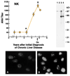
Fig. 2. Rise in ANA titer with malignancy
This patient developed HCC within 5 years after the diagnosis of chronic liver disease was made. Immunofluorescent ANA studies (frames A and B) were made at time points marked by asterisks and Western blotting (–4) at time points marked by vertical lines. Autoantibodies were already present before cancer detection (arrow) and additional new antibodies appeared with cancer (from Cancer 1993;71:26–35).
Fig. 3
Fig. 3. Expression of p62/IMP-II in HCC
Two HCC nodules were examined for expression of p62. (A) and (E) show hematoxylin–eosin staining of nodules and adjacent normal liver tissue. (B) and (F) show immunostaining of cancer nodules for p62 and absence of staining in adjacent normal liver. (C) and (G) show boxed areas enlarged: note cytoplasmic localization of p62 in cells in malignant nodules. (D) and (H) show DAPI staining of same areas as in (C) and (G) to demonstrate position of nuclei in the cells (from Am J Pathol 2001;159:945–953).
Fig. 4
Fig. 4. PCNA immunolocalization in non-synchronized HEp-2 tissue culture cells
The different patterns of nuclear staining corresponded cells in different phases of DNA synthesis, which were corroborated by separate thymidine uptake studies. Cells with little or no nuclear staining were in G1 or G2 phases of the cell cycle. Nucleolar and nucleoplasmic staining were cells in early S phase and cells with dense nucleoplasmic staining were in late S phase.
Fig. 5
Fig. 5. Construction of compound peptides of PCNA
The horizontal bar at the top represents full length PCNA with 261 amino acid residues. The shaded portions of this bar represent the regions that were selected for making compound peptides, with the arrowheads indicating the carboxy-terminus. Compound peptide 16-12 is a combination of peptide 106–112 covalently linked to 127–135 and so on. Compound peptide 15–52 is a combination of peptide 159–165 linked with an inverted 255–261, so that the latter has the carboxy-terminus joined to the carboxy-terminus of 159–165. Compound peptides 15–25 and 15–52 have the same composition of residues except that in the second half, the peptides are in reverse orientation (from J Biol Chem 1994;269:18,529–16,534).
Fig. 6
Fig. 6. Immunostaining of HEp-2 cells by 15–25 antiserum
Frame (A) shows immunostaining with 15–25 antiserum (fluorescein conjugate) followed by (B), double staining of same area with human anti-proliferating cell nuclear antigen antibody (rhodamine conjugate). (C) Another HEp-2 cell preparation after labeling with bromodeoxyuridine (BrdU) was immunostained with anti-15–25 (rhodamine conjugate) followed by double staining (D) with anti-BrdU (fluorescein conjugate). Arrows show cells in early S phase and arrowheads cells in mid or late S phase (from J Biol Chem 1994;269:18529–18534).

References

    1. Tan EM. Antinuclear antibodies: diagnostic markers for autoimmune diseases and probes for cell biology. Adv Immunol. 1989;44:93–151. - PubMed
    1. Tan EM. Interactions between autoimmunity and molecular and ell biology. Bridges between clinical and basic sciences. J Clin Invest. 1989;84:1–6. - PMC - PubMed
    1. Notkins A, Lernmark A. Autoimmune type 1 diabetes: resolved and unresolved issues. J Clin Invest. 2001;108:1247–1252. - PMC - PubMed
    1. Rapoport B, McLachlan SM. Thyroid autoimmunity. J Clin Invest. 2001;108:1253–1259. - PMC - PubMed
    1. Rose NR, Mackay IR. The Autoimmune Diseases. New York: Academic Press; 1998.

Publication types