. 2008;12(2):R42.
doi: 10.1186/cc6848. Epub 2008 Mar 25.

Circulating plasma factors induce tubular and glomerular alterations in septic burns patients

Affiliations

Circulating plasma factors induce tubular and glomerular alterations in septic burns patients

Filippo Mariano et al. Crit Care. 2008.

Abstract

Background: Severe burn is a systemic illness often complicated by sepsis. Kidney is one of the organs invariably affected, and proteinuria is a constant clinical finding. We studied the relationships between proteinuria and patient outcome, severity of renal dysfunction and systemic inflammatory state in burns patients who developed sepsis-associated acute renal failure (ARF). We then tested the hypothesis that plasma in these patients induces apoptosis and functional alterations that could account for proteinuria and severity of renal dysfunction in tubular cells and podocytes.

Methods: We studied the correlation between proteinuria and indexes of systemic inflammation or renal function prospectively in 19 severe burns patients with septic shock and ARF, and we evaluated the effect of plasma on apoptosis, polarity and functional alterations in cultured human tubular cells and podocytes. As controls, we collected plasma from 10 burns patients with septic shock but without ARF, 10 burns patients with septic shock and ARF, 10 non-burns patients with septic shock without ARF, 10 chronic uremic patients and 10 healthy volunteers.

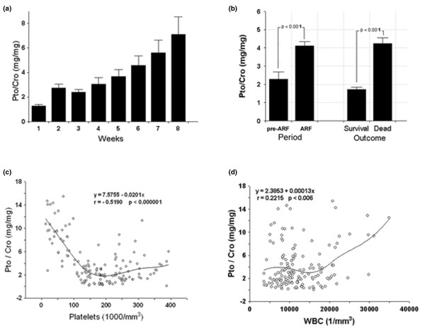
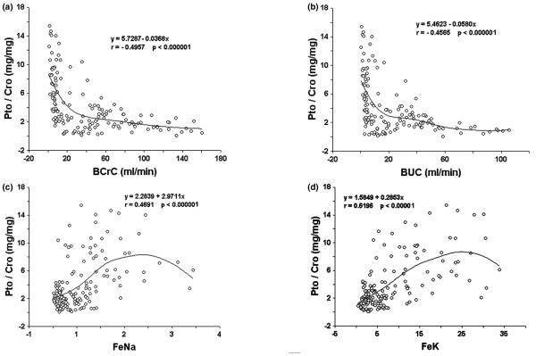
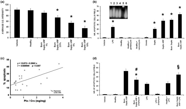
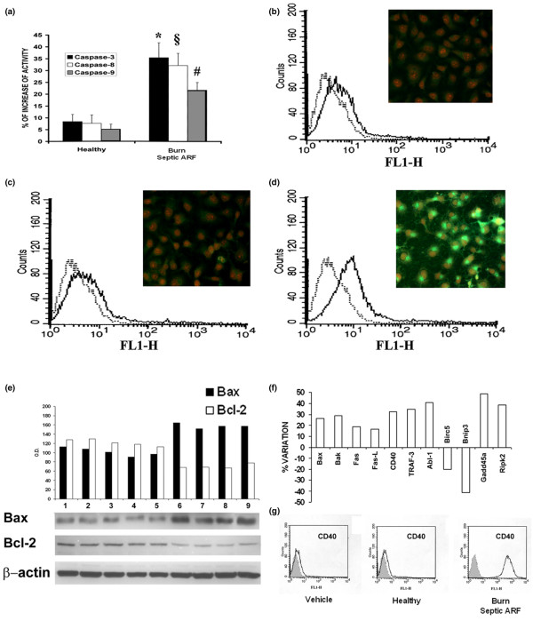
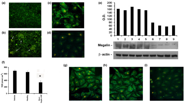
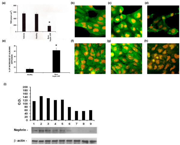
Results: Septic burns patients with ARF presented a severe proteinuria that correlated to outcome, glomerular (creatinine/urea clearance) and tubular (fractional excretion of sodium and potassium) functional impairment and systemic inflammation (white blood cell (WBC) and platelet counts). Plasma from these patients induced a pro-apoptotic effect in tubular cells and podocytes that correlated with the extent of proteinuria. Plasma-induced apoptosis was significantly higher in septic severe burns patients with ARF with respect to those without ARF or with septic shock without burns. Moreover, plasma from septic burns patients induced an alteration of polarity in tubular cells, as well as reduced expression of the tight junction protein ZO-1 and of the endocytic receptor megalin. In podocytes, plasma from septic burns patients increased permeability to albumin and decreased the expression of the slit diaphragm protein nephrin.

Conclusion: Plasma from burns patients with sepsis-associated ARF contains factors that affect the function and survival of tubular cells and podocytes. These factors are likely to be involved in the pathogenesis of acute tubular injury and proteinuria, which is a negative prognostic factor and an index of renal involvement in the systemic inflammatory reaction.


Figures

Figure 1
Figure 1
Proteinuria correlates with patient outcome and with markers of systemic inflammation. (a) Proteinuria expressed as proteinuria/creatininuria ratio (Pto/Cro, mg/mg) in the weeks following patient admission. Data are given as weekly average of daily values. Only non-oliguric patients were included and the number of patients for each week was: week 1, n = 19; week 2, n = 19; week 3, n = 17; week 4, n = 15; week 5, n = 10; week 6, n = 7; week 7, n = 6; week 8, n = 5. (b) Overall mean proteinuria in pre-acute renal failure (ARF) vs ARF periods and in deceased vs surviving patients. (c, d) Relationship between Pto/Cro and indexes of systemic inflammatory state in ARF period. Pto/Cro negatively correlated with platelet count (c) and positively with white blood cell (WBC) count (d). Student t test and linear regression analysis were performed where appropriate. Data for different parameters are also shown as minimal square fitting curves.
Figure 2
Figure 2
Correlation among proteinuria and indexes of glomerular and tubular function in acute renal failure (ARF) period. (a, b) Significant negative relationship between proteinuria/creatininuria ratio (Pto/Cro, mg/mg) and blood creatinine clearance (BCrC, ml/min, (a)) and blood urea clearance (BUC, ml/min, (b)). (c, d) Significant positive relationship between Pto/Cro and fractional excretion of sodium (FeNa, (c)) and potassium (FeK, (d)). A statistical linear regression test analysis was performed. Data for different parameters are also shown as minimal square fitting curves.
Figure 3
Figure 3
Pro-apoptotic effect of burns septic acute renal failure (ARF) group plasma on tubular cells and correlation with proteinuria. (a) Plasma from burns septic ARF group patients induced a dose-dependent decrease of tubular cell viability (XTT-based assay, n = 19, *p < 0.05 burns septic ARF group plasma 2.5%, 5% or 10% vs control healthy plasma). (b) Burns septic ARF group plasma (5%, 48 h of incubation, n = 19) induced a significant increase in tubular apoptosis (terminal uridine deoxynucleotidyl transferase dUTP nick-end labeling (TUNEL) assay, *p < 0.05 burns septic ARF group plasma vs control healthy plasma, healthy plasma + gentamicin, healthy plasma + vancomycin or uremic plasma). Gentamicin (2 μg/ml) or vancomycin (10 μg/ml) was added to control healthy plasma in selected experiments (n = 10). A significant increase of tubular apoptosis with a maximal effect with burns septic ARF group plasma (*p < 0.05 septic, septic ARF or burns septic vs all controls) was observed. LPS (30 ng/ml) was used as positive experimental control. (b) inset, typical DNA fragmentation of apoptotic tubular cells (burns septic ARF group plasma in lanes 1–4, positive control 30 ng/ml LPS in lane 6, negative control healthy plasma in lane 5). (c) Correlation between the percentage of tubular apoptosis induced by burns septic ARF group plasma (TUNEL assay) and Pto/Cro of the enrolled patients (n = 19). (d) Significant reduction of tubular apoptosis (TUNEL assay) after addition of 5 μg/ml polymyxin B (#p < 0.05 burns septic ARF group + polymyxin vs burns septic ARF group, n = 19). Lipopolysaccharide (LPS; 30 ng/ml) was used as internal control. Polymyxin B pre-treatment did not completely suppress plasma-induced apoptosis (*p < 0.05 burns septic ARF group + polymyxin vs control healthy plasma). Burns septic ARF group plasma but not control healthy plasma enhanced LPS-induced tubular apoptosis (§p < 0.05 burns septic ARF group plasma + LPS vs control healthy plasma + LPS). Values in (a), (b) and (d) are expressed as averages ± standard error (SE). Each plasma was tested in triplicate. Analysis of variance (ANOVA) with Newman-Keuls multi-comparison test was performed. Linear regression analysis was performed in (c).
Figure 4
Figure 4
Burns septic ARF group plasma activated caspases, up-regulated Fas/CD40 and modulated Bax/Bcl-2 in tubular cells. (a) Significant increased activities of caspases 3, 8 and 9 on tubular cells incubated for 48 h with burns septic acute renal failure (ARF) group plasma (n = 19) in comparison to control healthy plasma (n = 10). All plasma samples were tested in triplicate. Student t test was performed: (p < 0.05 *caspase-3, §caspase-8 and #caspase-9 activities of burns septic ARF group vs control healthy plasma). (b-d) Representative images of fluorescence-activated cell sorting (FACS) and immunofluorescence (insets) analysis of Fas (CD95) expression on tubular cell surface after exposure to different stimuli. With respect to vehicle alone (b) or control healthy plasma (c), burns septic ARF group plasma induced a marked up-regulation of Fas (d) (magnification × 400, nuclei counterstained with 1 μg/ml propidium iodide). Similar results were obtained with all tested plasma. (e) Up-regulation of the pro-apoptotic protein Bax and down-regulation of the antiapoptotic protein Bcl-2 in representative Western blot analysis on lysates of tubular cells (vehicle alone in lane 1, control healthy plasma in lanes 2–5, burns septic ARF group plasma in lanes 6–9) and related densitometric analysis. Beta-actin was used as reference for protein loading. (f) Percentage variation of expression of genes involved in apoptosis of tubular cells exposed to burns septic ARF group plasma. Tubular cells showed an increased expression of genes related to receptor-mediated (Fas, Fas-Ligand) and mitochondrial apoptotic pathways (Bax, Bak), of the co-stimulatory molecule CD40, of the CD40-transducer protein TRAF-3 and of the positive regulator of nuclear factor (NF)-κB activator RIPK2. Results are given as ratio between densitometric analyses of gene expression in tubular cells exposed to burns septic ARF group plasma with respect to control healthy plasma. House-keeping genes (beta-actin, GAPDH) were used as reference for densitometric analysis. Three experiments were performed with similar results. These data are feely available from the ArrayExpress databank of the European Bioinformatics Institute (see Materials and methods). Representative FACS analysis of CD40 expression was performed on unstimulated tubular cells (Vehicle), or in the presence of control healthy plasma, or burns septic ARF group plasma. Similar results were obtained with all tested burns septic ARF group plasma. The grey areas show the isotypic control.
Figure 5
Figure 5
Burns septic acute renal failure (ARF) group plasma altered cytoskeleton, megalin and tight junction expression and polarity in tubular cells. (a, b) Representative micrographs showing normal distribution ((a) control healthy plasma) and marked re-arrangement of cytoskeleton actin on tubular cells exposed for 48 h to burns septic ARF group plasma (b) with formation of "heaps" (white arrows) visible via ultraviolet (UV) light microscopy after staining with fluorescein isothiocyanate (FITC)-conjugated phalloidin (magnification × 100). (c, d) Representative immunofluorescence of megalin expression on tubular cells incubated with control healthy plasma (c) and its down-regulation in presence of burns septic ARF group plasma (d) (magnification × 400, nuclei counterstained with 1 μg/ml propidium iodide). Similar results were observed with all tested plasma samples. (e) Representative Western blot analysis of megalin expression on tubular cells (vehicle alone in lane 1, control healthy plasma in lanes 2–5, burns septic ARF group plasma in lanes 6–9) and densitometric analysis. Beta-actin was used as reference for protein loading. (f) Significant loss of polarity of tubular cells evaluated by the decrease of trans-epithelial resistance (TER) normalized for the membrane area after incubation with burns septic ARF group plasma for 12 h (*p < 0.05 burns septic ARF group vs control healthy plasma). (g-i) Representative micrographs showing the expression of the tight junction protein ZO-1 on unstimulated tubular cells (g), in the presence of control healthy plasma (h) and its down-regulation after incubation with burns septic ARF group plasma (i) (magnification × 400, nuclei counterstained with 1 μg/ml propidium iodide). Similar results were obtained with all tested plasma samples. Values in (f) are expressed as averages ± standard error (SE). Each plasma sample was tested in triplicate. Analysis of variance (ANOVA) with Newman-Keuls multi-comparison test was performed.
Figure 6
Figure 6
Burns septic acute renal failure (ARF) group plasma altered polarity, permeability to albumin and nephrin expression on podocytes. (a) Significant variation of trans-epithelial resistance (TER) after exposure to burns septic ARF group plasma (*p < 0.05 burns septic ARF group vs control healthy plasma). (b-d) Representative immunofluorescence images of the altered distribution of the intermediate filament protein nestin in presence of burns septic plasma (d), not detectable after incubation with vehicle alone (b) or control healthy plasma (c) (magnification × 400, nuclei counterstained with 1 μg/ml propidium iodide). After burns septic plasma challenge, nestin lost its typical diffuse distribution and was localized in the sub-membrane spaces forming several "rings" (d). (e) Significant increased diffusion of albumin across podocyte monolayers exposed to burns septic ARF group plasma (*p < 0.05 burns septic plasma vs control healthy plasma). (f-h) Representative immunofluorescence images of the slit diaphragm protein nephrin after exposure to vehicle alone (f), control healthy plasma (g) and burns septic ARF group plasma (h). Burns septic ARF group plasma induced a diffuse loss of nephrin (magnification × 400, nuclei counterstained with 1 μg/ml propidium iodide). (i) Representative Western blot analysis of podocyte nephrin expression (vehicle alone in lane 1, control healthy plasma in lanes 2–5, burns septic ARF group plasma in lanes 6–9) and related densitometric analysis. Beta-actin was used as reference for protein loading. Values in (a) and (e) are expressed as averages ± standard error (SE). Each plasma was tested in triplicate. Analysis of variance (ANOVA) with Newman-Keuls multi-comparison test was performed.

Comment in

Similar articles

Cited by

References

    1. Schrier RW, Wang W. Acute renal failure and sepsis. N Engl J Med. 2004;351:159–169. doi: 10.1056/NEJMra032401. - DOI - PubMed
    1. Hotchkiss RS, Swanson PE, Cobb JP, Jacobson A, Buchman TG, Karl IE. Apoptosis in lymphoid and parenchymal cells during sepsis: findings in normal and T- and B-cell-deficient mice. Crit Care Med. 1997;25:1298–1307. doi: 10.1097/00003246-199708000-00015. - DOI - PubMed
    1. Camussi G, Mariano F, Biancone L, De Martino A, Bussolati B, Montrucchio G, Tobias PS. Lipopolysaccharide binding protein and CD14 modulate the synthesis of platelet-activating factor by human monocytes and mesangial and endothelial cells stimulated with lipopolysaccharide. J Immunol. 1995;155:316–324. - PubMed
    1. Cunningham PN, Dyanov HM, Park P, Wang J, Newell KA, Quigg RJ. Acute renal failure in endotoxemia is caused by TNF acting directly on TNF receptor-1 in kidney. J Immunol. 2002;168:5817–5823. - PubMed
    1. Wan L, Bellomo R, Di Giantomasso D, Ronco C. The pathogenesis of septic acute renal failure. Curr Opin Crit Care. 2002;9:496–502. doi: 10.1097/00075198-200312000-00006. - DOI - PubMed

Publication types

MeSH terms