Comparative Study
. 2008 Jul 1;17(13):1946-55.
doi: 10.1093/hmg/ddn092. Epub 2008 Mar 25.

Lysosomal proliferation and distal degeneration in motor neurons expressing the G59S mutation in the p150Glued subunit of dynactin

Affiliations
Comparative Study

Lysosomal proliferation and distal degeneration in motor neurons expressing the G59S mutation in the p150Glued subunit of dynactin

Erica S Chevalier-Larsen et al. Hum Mol Genet. .

Abstract

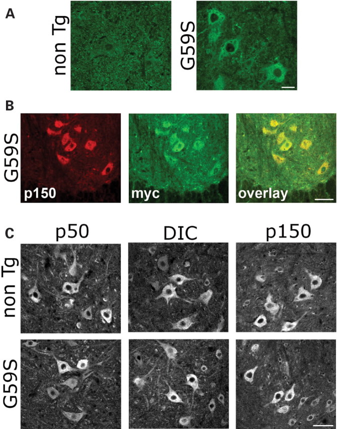
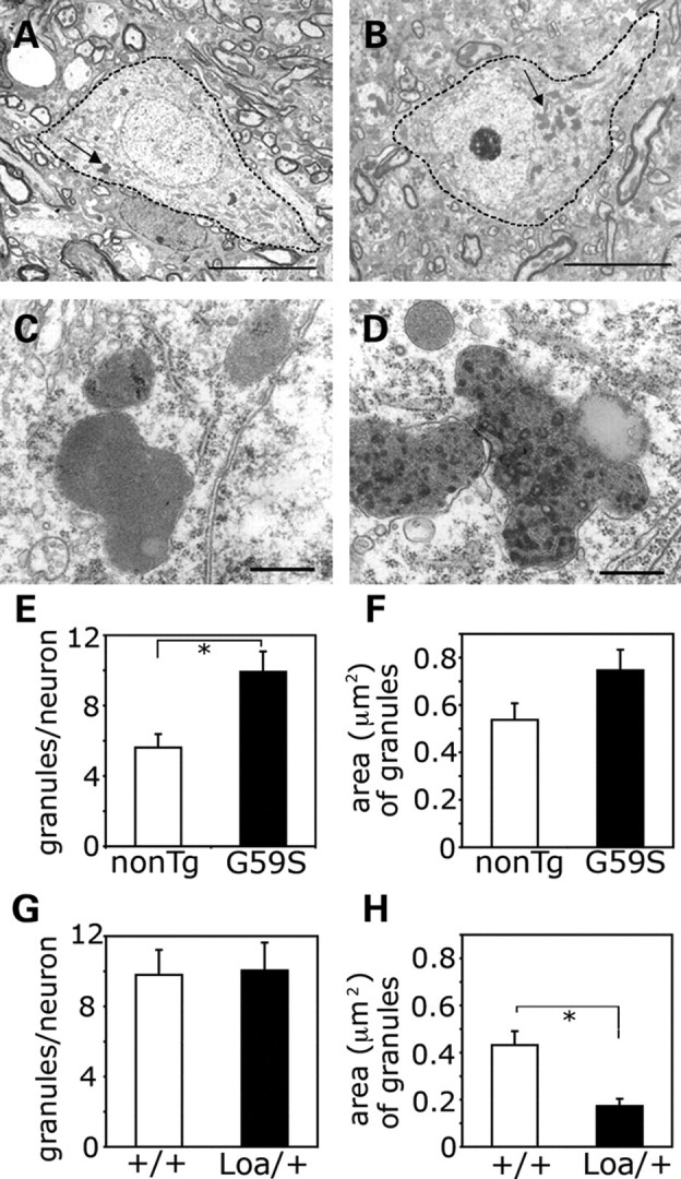
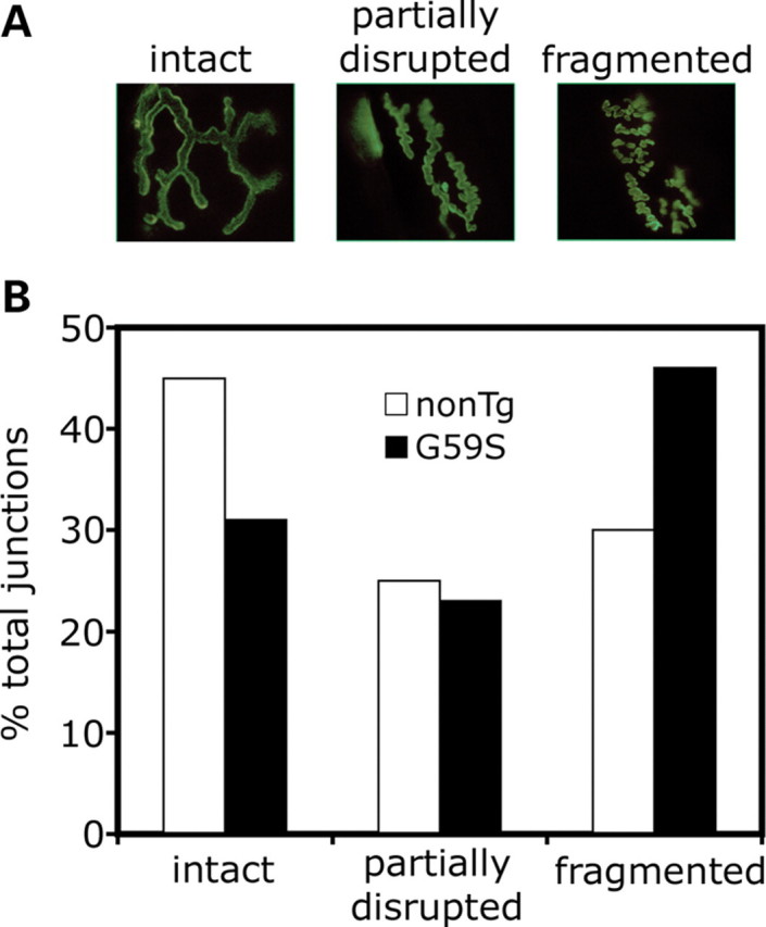
An increasing number of neurodegenerative diseases are being linked to mutations in genes encoding proteins required for axonal transport and intracellular trafficking. A mutation in p150(Glued), a component of the cytoplasmic dynein/dynactin microtubule motor complex, results in the human neurodegenerative disease distal spinal and bulbar muscular atrophy (dSBMA). We have developed a transgenic mouse model of dSBMA; these mice exhibit late-onset, slowly progressive muscle weakness but do not have a shortened lifespan, consistent with the human phenotype. Examination of motor neurons from the transgenic model reveals the proliferation of enlarged tertiary lysosomes and lipofuscin granules, indicating significant alterations in the cellular degradative pathway. In addition, we observe deficits in axonal caliber and neuromuscular junction (NMJ) integrity, indicating distal degeneration of motor neurons. However, sciatic nerve ligation studies reveal that inhibition of axonal transport is not evident in this model. Together, these data suggest that mutant p150(Glued) causes neurodegeneration in the absence of significant changes in axonal transport, and therefore other functions of dynein/dynactin, such as trafficking in the degradative pathway and stabilization of the NMJ are likely to be critical in maintaining the health of motor neurons.


Figures

Figure 1.
Figure 1.
Generation and characterization of a transgenic mouse expressing G59S p150Glued. (A) Top: schematic of the transgene construct, including the Thy1.2 promoter to drive neuron-specific expression, the p150Glued coding region with the G59S point mutation, and a C-terminal myc tag. Bottom: screening of stably integrated lines (M16, M20 and M22) for expression of the transgene in spinal cord and brain using an anti-myc antibody, in comparison with a non-transgenic line and positive and negative controls for the myc antibody (extracts of Cos7 cells transfected with p150G59S-myc-tagged plasmid, or untransfected cells, respectively). (B) Quantitative western blotting of spinal cord extracts from non-transgenic and TgG59S mice, probed with a monoclonal antibody to p150Glued that recognizes only the endogenous polypeptide, and a polyclonal antibody that recognizes both the endogenous and G59S forms of p150Glued. The blots were also probed with antibodies to the myc-tag of the transgene, dynein (DIC), and actin. (C) Brain lysates from non-transgenic control mice (top) and TgG59S mice (bottom) were fractionated on sucrose density gradients, and then analyzed by SDS–PAGE and western blot, using antibodies to dynein (DHC, DIC), dynactin (p150, p50), the myc tag, and actin. Dynein and dynactin co-sediment at ∼20S from both control and TgG59S mice, indicating that expression of the transgene does not disrupt endogenous dynein or dynactin. (D) TgG59S female mice show mild, progressive declines in forelimb grip strength in comparison with age-matched non-transgenic littermate controls. Grip strength was measured monthly starting at 2 months of age and continuing until 18 months of age. Asterisks indicate a statistical difference determined by t-test at that time point, with P < 0.05 (n ≥ 12 per genotype); error bars represent SEM.
Figure 2.
Figure 2.
Expression of the G59S p150Glued transgene in this model does not significantly inhibit axonal transport in the TgG59S model. Sciatic nerves were ligated and accumulation of motor proteins proximal and distal to the block was analyzed to compare axonal transport in TgG59S and non-transgenic control mice. (A) Immunoblots of 2 mm nerve segments immediately proximal and distal to the site of ligature following a 2 h ligation were probed with antibodies to dynein (DHC), dynactin (p150Glued), and kinesin; glyceraldehyde 3-phosphate dehydrogenase (GAPDH) was used as a loading control. In both transgenic and non-transgenic mice, kinesin accumulates proximal to the block, indicating efficient ligation of the nerve. No significant differences were observed in the accumulation of dynein and dynactin distal to the block in transgenic as compared with non-transgenic mice, when corrected for differences in loading. (B) Average motor protein accumulation proximal and distal to the ligation for each genotype was quantitated as signal intensity relative to a GAPDH loading control. Differences in accumulation of dynein or dynactin distal to the block in TgG59S relative to non-transgenic controls were not statistically different (t-test, P > 0.25, n = 6 mice per genotype; error bars represent SEM).
Figure 3.
Figure 3.
The G59S p150Glued transgene is expressed in motor neurons of the spinal cord. (A) Spinal cord sections from a 9-month-old TgG59S mouse and an age-matched littermate control were stained with an antibody to the myc-tag to visualize expression of the transgene in motor neurons. No significant staining is seen in the age-matched non-transgenic (non-Tg) control. (B) A section from the ventral horn of the lumbar spinal cord from a TgG59S mouse was double-stained with antibodies to p150Glued (red) and the myc tag (green). The merged image (overlay) shows that these motor neurons are expressing the p150-G59S-myc transgene. Scale bar = 50 μm. (C) Lumbar spinal cord sections from non-transgenic (non-Tg) and TgG59S transgenic (G59S) mice were stained with antibodies to the dynamitin (p50) and p150Glued (p150) subunits of dynactin and the DIC subunit of cytoplasmic dynein. Scale bar = 50 μm.
Figure 4.
Figure 4.
Electron microscope analysis of cervical spinal cord reveals the proliferation of degradative organelles, including lysosomes and lipofuscin granules, in TgG59S mice. (A, B) Micrographs of motor neurons from age-matched non-transgenic (A) and TgG59S (B) mice demonstrate the proliferation of degradative organelles in the perinuclear region of the cell in transgenic mice relative to non-transgenic controls. Dashed lines indicate the cell membrane of the motor neuron; arrows point to degradative organelles; scale bar = 10 µm. (C, D) In higher resolution images these organelles can be identified as lipofuscin granules; these organelles are both more numerous and more heterogeneous in motor neurons of TgG59S mice (D) compared with age-matched non-transgenic controls (C); scale bar = 500 nm. (E) Quantitative analysis indicates a significant increase in the number of lipofuscin granules in spinal motor neurons from TgG59S mice as compared with non-transgenic age-matched controls (t-test, P < 0.002, n ≥ 20 cells, three mice per genotype). (F) Average lipofuscin granule size is also increased in TgG59S mice when compared with age-matched controls although not significantly (t-test, P < 0.07, n ≥ 20 cells, three mice per genotype). (G) Quantitative analysis of lipofuscin granules in motor neurons from Loa/+ heterozygous mice in comparison with +/+ littermate control mice indicates that the dynein mutation does not induce an upregulation in the number of granules per cell (t-test, P > 0.9, n ≥ 20 cells, three mice per genotype). (H) The average size of lipofuscin granules in Loa/+ mice is decreased relative to control mice (t-test, P < 0.001, n ≥ 20 cells, three mice per genotype). Error bars in all graphs represent SEM.
Figure 5.
Figure 5.
Reduced axonal caliber of motor neurons from TgG59S mice. (A and B) Cross-sectional images of L5 ventral roots from 18-month-old TgG59S mice (G59S) and age-matched control mice (non-Tg). There is a clear decrease in axonal caliber in TgG59S mice relative to controls. (C) Relative frequency distributions for axonal calibers from the L5 ventral root of TgG59S mice as compared with age-matched non-transgenic controls (K–S test, P « 0.001, n = 3 mice per genotype). (D) The percentage of demyelinated axons is similar between TgG59S mice and controls (t-test, P > 0.25, n = 3 mice per genotype); the percentage of remyelinated axons is decreased, but not significantly (t-test, P > 0.1, n = 3 mice per genotype).
Figure 6.
Figure 6.
NMJ morphology is disrupted in TgG59S mice. (A) Representative pictures of the neuromuscular junctions of soleus muscle from 18-month-old TgG59S and controls visualized by immunostaining the presynaptic terminal with antibodies to neurofilaments and SV2 (both in green). Junctions were scored as intact, partially disrupted, or fragmented, as shown. (B) TgG59S mice have a greater proportion of fragmented junctions and fewer intact junctions than age-matched non-transgenic controls.
Figure 7.
Figure 7.
Comparison of the pathological hallmarks of neurodegeneration in multiple mouse models. Various pathological features associated with motor neuron degenerative disease are listed in boxes. Experimental data on mouse models with disruptions in either dynein or dynactin are summarized; factors that have been observed in the various models are listed in green text; whereas mouse models experimentally shown to lack a particular feature are listed in red. Supporting data for this model was gathered from multiple references (,–7,16).

References

    1. Chevalier-Larsen E., Holzbaur E.L. Axonal transport and neurodegenerative disease. Biochim. Biophys. Acta. 2006;1762:1094–1108. - PubMed
    1. Puls I., Jonnakuty C., LaMonte B.H., Holzbaur E.L., Tokito M., Mann E., Floeter M.K., Bidus K., Drayna D., Oh S.J., et al. Mutant dynactin in motor neuron disease. Nat. Genet. 2003;33:455–456. - PubMed
    1. Waterman-Storer C.M., Karki S., Holzbaur E.L. The p150Glued component of the dynactin complex binds to both microtubules and the actin-related protein centractin (Arp-1) Proc. Natl Acad. Sci. USA. 1995;92:1634–1638. - PMC - PubMed
    1. Levy J.R., Sumner C.J., Caviston J.P., Tokito M.K., Ranganathan S., Ligon L.A., Wallace K.E., LaMonte B.H., Harmison G.G., Puls I., et al. A motor neuron disease-associated mutation in p150Glued perturbs dynactin function and induces protein aggregation. J. Cell Biol. 2006;172:733–745. - PMC - PubMed
    1. Puls I., Oh S.J., Sumner C.J., Wallace K.E., Floeter M.K., Mann E.A., Kennedy W.R., Wendelschafer-Crabb G., Vortmeyer A., Powers R., et al. Distal spinal and bulbar muscular atrophy caused by dynactin mutation. Ann. Neurol. 2005;57:687–694. - PMC - PubMed

Publication types

MeSH terms