. 2008 Apr 1;105(13):5207-12.
doi: 10.1073/pnas.0711558105. Epub 2008 Mar 25.

The vitamin D receptor is required for iNKT cell development

Affiliations

The vitamin D receptor is required for iNKT cell development

Sanhong Yu et al. Proc Natl Acad Sci U S A. .

Abstract

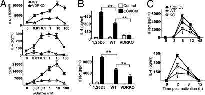
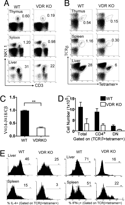
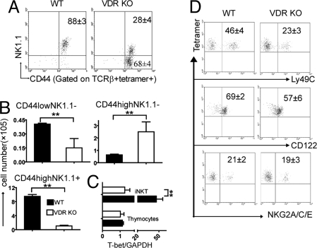
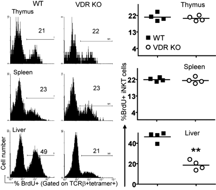
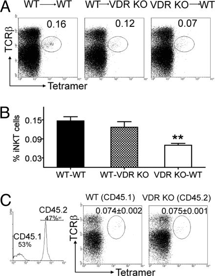
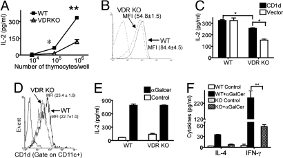
CD1d-reactive natural killer T (NKT) cells with an invariant T cell receptor Valpha14 rearrangement are a unique subset of lymphocytes, which play important roles in immune regulation, tumor surveillance, and host defense against pathogens. Vitamin D is a nutrient/hormone that has been shown to regulate conventional T cell responses but not T cell development. The data show that expression of the vitamin D receptor (VDR) is required for normal development and function of iNKT cells. The iNKT cells from VDR KO mice are intrinsically defective and lack T-bet expression. VDR KO iNKT cells fail to express NK1.1, although they express normal levels of CD122. Extrinsic factors that impact iNKT cell development and function in VDR KO mice include a failure of the liver to support homeostatic proliferation and reduced thymic expression of CD1d and other factors important for optimal antigen presentation in the thymus. In addition, VDR KO iNKT cells were intrinsically defective even when WT antigen-presenting cells were used to stimulate them.


Conflict of interest statement

The authors declare no conflict of interest.

Figures

Fig. 1.
Fig. 1.
iNKT cells from VDR KO mice are hyporesponsive. (A) Splenocytes from WT and VDR KO mice were stimulated with different concentrations of αGalCer in vitro. IFN-γ, IL-4, and proliferation were measured. Graphs represent mean ± SD from triplicate samples. Data are one representative experiment of three. (B) IL-4 and IFN-γ production by splenocytes stimulated with 10 nM αGalCer. The splenocytes were from 1,25(OH)2D3-fed WT (1,25D3), WT, and VDR KO mice. Data are from the mean ± SEM of three experiments. **, P < 0.001. (C ) Serum cytokine production in WT and VDR KO mice induced by systemic administration of αGalCer. Levels of IFN-γ and IL-4 in the serum were determined at different times after injection (n = 15 per group, *, P < 0.05).
Fig. 2.
Fig. 2.
Reduced NKT cell development in VDR KO mice. (A) Dot plots showing percentage of NK1.1 and CD3-positive NKT cells in VDR KO and WT controls. Data show one representative of 25 mice per group. Numbers indicate percentage of NK1.1 and CD3 double-positive cells. Fig. S2A shows the means ± SEM for NK1.1 and CD3 double staining. (B) Dot plot showing iNKT cells (TCRβ and CD1d-αGalCer double-positive). Fig. S3D shows the TCRβ empty CD1d control staining. Data show one representative of 20 mice per group. Numbers indicate percentage of iNKT cells in the circled gate. (C ) Real-time PCR analysis of the TCR Vα14-Jα18 expression in WT and VDR KO thymocytes. Expression of the TCR constant β chain was used as an amplification control. Data are the mean of three independent experiments ± SEM. **, P < 0.001. (D) The absolute number of total iNKT, CD4+ iNKT, and DN iNKT (TCRβ and tetramer double-positive) cells in the spleen were compared between WT and VDR KO mice (n = 15). Fig. S3B shows the isotype control for CD4 staining. (E) Frequency of cytokine-producing iNKT cells in WT and VDR KO mice. Mice were injected with αGalCer in vivo followed by intracellular cytokine staining ex vivo as described in Materials and Methods. Histograms show production of IFN-γ and IL-4 by iNKT cells. Data from one representative of 10 mice. Fig. S1C shows the means ± SEM for the IL-4 and IFN-γ responses. Fig. S3A shows the isotype control staining for both IL-4 and IFN-γ.
Fig. 3.
Fig. 3.
VDR KO iNKT cells fail to mature fully. (A) Dot plots showing expression of CD44 and NK1.1 on TCRβ and CD1d-αGalCer tetramer double-positive thymocytes. Data from one representative of 10 mice is shown. (B) Absolute numbers of CD44lowNK1.1, CD44highNK1.1, and CD44highNK1.1+ iNKT cells in the thymus of WT and VDR KO mice (n = 10, **, P < 0.001). (C ) Real-time PCR analysis of T-bet expression in thymocytes and thymic iNKT cells from WT and VDR KO mice. Expression of GAPDH was used as an amplification control. Data are the mean of two independent experiments ± SEM. **, P < 0.001. (D) Tetramer-positive thymocytes were analyzed for Ly49C, CD122, and NKG2A/C/E expression. Data from one representative of six mice is shown.
Fig. 4.
Fig. 4.
Reduced iNKT cell homeostatic proliferation in the liver of VDR KO mice. BrdU incorporation by iNKT cells from WT and VDR KO mice. Fig. S3C shows the BrDU isotype control staining. Histograms of one representative of four mice. (Right) Individual values and the mean (line) of the data. **, P < 0.001.
Fig. 5.
Fig. 5.
iNKT cell development requires VDR expression in the hematopoetic compartment. (A) Reciprocal BM transplants were done by using WT (CD45.1) and VDR KO (CD45.2) mice (donor BM → recipient). The success of the transplant was determined by staining for donor cells in the recipient mice. Percentage of donor iNKT cells in the thymus is shown. Data shown is one representative of four mice per group and two independent experiments. Fig. S4A shows the percentage of donor iNKT cells in liver and spleen of the recipient mice (mean ± SEM of eight mice). (B) Pooled data showing the % of donor iNKT cells recovered in the thymus of reciprocal BM transplantation experiments. Data are the mean ± SEM of eight mice. **, P < 0.01. (C ) Competitive BM chimeras were generated by using a 1:1 ratio of WT CD45.1 and VDR KO CD45.2 BM and WT CD45.1 recipients. Thymocyte chimerism was determined by flow cytometry (Left) and shown to be 53% of WT CD45.1 origin and 47% of VDR KO CD45.2 origin. Fig. S4B shows the percentage of CD45.1 and CD45.2 iNKT cells in the liver and spleen of recipient mice. The data shown is from one representative of five mice (mean ± SEM) and one of two experiments.
Fig. 6.
Fig. 6.
Low CD1d expression and reduced antigen presentation in the thymus of VDR KO mice. (A) Equal numbers of the CD1d-restricted NKT hybridoma cell were incubated in the presence of different concentrations of thymocytes from WT and VDR KO mice and IL-2 production by the NKT cell hybridoma was measured. Results shown are from one representative of four independent experiments. *, P < 0.05; **, P < 0.001. (B) Thymocytes isolated from WT and VDR KO mice were analyzed by flow cytometry for CD1d expression. Mean fluorescent intensity (MFI) ± SEM. Data shown are from one representative of 15 mice. (C) Thymocytes from WT and VDR KO mice were infected with a CD1d-expressing retrovirus or empty vector and used to stimulate the NKT cell hybridoma for IL-2 production. Graph represents the mean ± SD from triplicate samples. Data shown are one individual experiment of two. *, P < 0.05. (D) CD1d expression on purified VDR KO and WT DC (MFI ± SEM). (E) Purified DC from WT and VDR KO spleens were pulsed with αGalCer or media (Control) and cocultured with CD1d-restricted hybridoma cells for IL-2 production. Graph represents the mean ± SD from triplicate samples. Data shown are one representative experiment of two. (F) iNKT cells from WT and VDR KO (KO) mice were purified and cultured with Rag KO splenocytes in media only (Control) or with αGalCer. Graph represents the mean ± SD from triplicate samples. **, P < 0.001.

References

    1. Godfrey DI, Hammond KJ, Poulton LD, Smyth MJ, Baxter AG. NKT cells: Facts, functions and fallacies. Immunol Today. 2000;21:573–583. - PubMed
    1. Bendelac A, Bonneville M, Kearney JF. Autoreactivity by design: Innate B and T lymphocytes. Nat Rev Immunol. 2001;1:177–186. - PubMed
    1. Gumperz J, Miyake S, Yamamura T, Brenner MB. Functionally distinct subsets of CD1d-restricted natural killer T cells revealed by CD1d tetramer staining. J Exp Med. 2002;195:625–636. - PMC - PubMed
    1. Taniguchi M, Harada M, Kojo S, Nakayama T, Wakao H. The regulatory role of Valpha14 NKT cells in innate and acquired immune response. Annu Rev Immunol. 2003;21:483–513. - PubMed
    1. Bendelac A, Rivera MN, Park SH, Roark JH. Mouse CD1-specific NK1 T cells: Development, specificity, and function. Annu Rev Immunol. 1997;15:535–562. - PubMed

Publication types

MeSH terms