. 2008 Mar;20(3):614-34.
doi: 10.1105/tpc.106.047423. Epub 2008 Mar 25.

Exclusion of a proton ATPase from the apical membrane is associated with cell polarity and tip growth in Nicotiana tabacum pollen tubes

Affiliations

Exclusion of a proton ATPase from the apical membrane is associated with cell polarity and tip growth in Nicotiana tabacum pollen tubes

Ana C Certal et al. Plant Cell. 2008 Mar.

Abstract

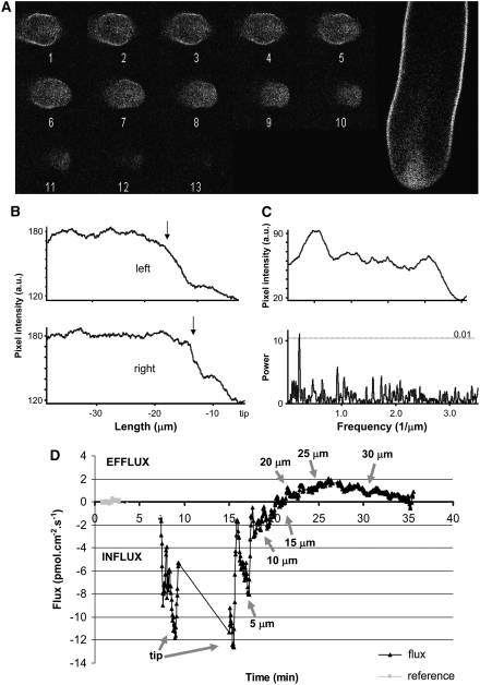
Polarized growth in pollen tubes results from exocytosis at the tip and is associated with conspicuous polarization of Ca(2+), H(+), K(+), and Cl(-) -fluxes. Here, we show that cell polarity in Nicotiana tabacum pollen is associated with the exclusion of a novel pollen-specific H(+)-ATPase, Nt AHA, from the growing apex. Nt AHA colocalizes with extracellular H(+) effluxes, which revert to influxes where Nt AHA is absent. Fluorescence recovery after photobleaching analysis showed that Nt AHA moves toward the apex of growing pollen tubes, suggesting that the major mechanism of insertion is not through apical exocytosis. Nt AHA mRNA is also excluded from the tip, suggesting a mechanism of polarization acting at the level of translation. Localized applications of the cation ionophore gramicidin A had no effect where Nt AHA was present but acidified the cytosol and induced reorientation of the pollen tube where Nt AHA was absent. Transgenic pollen overexpressing Nt AHA-GFP developed abnormal callose plugs accompanied by abnormal H(+) flux profiles. Furthermore, there is no net flux of H(+) in defined patches of membrane where callose plugs are to be formed. Taken together, our results suggest that proton dynamics may underlie basic mechanisms of polarity and spatial regulation in growing pollen tubes.


Figures

Figure 1.
Figure 1.
Localization of Nt AHA-GFP in Transformed Pollen Tubes. (A) Control pollen tube transformed with LAT52-NcoI-GFP plasmid. (B) Longitudinal optical section of the middle part of a pollen tube showing labeling in the membrane. (C) Plasmolyzed pollen grain with labeled membrane detached from the autofluorescent exine. (D) Pollen tube labeling upon osmotic shock with strands of plasma membrane linking the plasmolysed cytoplasm (arrows). g, pollen grain. (E) High-contrast two-photon optical section of a pollen tube showing the V-shape intracellular labeling region at the tip. (F) Nongrowing pollen tube showing the membrane labeling pattern but lacking the tip-localized intracellular labeling. Bars = 5 μm.
Figure 2.
Figure 2.
Fluorescence Profiling of Nt AHA-GFP Labeling. (A) Sequential transversal planes (1 μm apart) of the tip of a growing Nt AHA-GFP transgenic pollen tube; plasma membrane Nt AHA-GFP labeling decreases toward the apex (plane 13) where only intracellular labeling is detected. (B) Membrane labeling profile with fluorescence decay at ∼15 μm from the tip (arrows) at both sides of a pollen tube and almost no labeling detected at the very tip. a.u., arbitrary fluorescence intensity units. (C) Spectral analysis of a labeling profile where a fluctuation pattern in the fluorescence was observed; the bottom graph shows a significant frequency peak corresponding to a spatial period of ∼10 μm. (D) H+ flux profile of a LAT52:NtAHA-GFP tube showing that the spatial distribution of fluxes matches the fluorescence profile for these proton pumps.
Figure 3.
Figure 3.
FRAP in Nt AHA-GFP–Labeled Pollen Tubes. (A) Image showing the typical fluorescence hole produced after bleaching, also depicting pollen tube orientation. (B) Graphical depiction of the movement of the mathematical centroid of a square box area containing the bleached patch of membrane throughout fluorescence recovery. (C) Images representing the first (t = 0 s) and last (t = 320 s) frames of the analyzed time sequence with the centroid represented by asterisks (algorithms for centroid calculation are the ones used in the track object function of the software package Metamorph).
Figure 4.
Figure 4.
Nt AHA mRNA Localization in Pollen Tubes. (A) Pollen tube hybridized with antisense Probe 3 showing no labeling at the tip region. The top panel shows the raw two-photon image, and the bottom panel shows the respective pseudocolor image. (B) Pollen tube with an unpreserved intracellular structure (vacuole present at the tip, V) hybridized with antisense Probe 3 and showing labeling at the tip region. (C) Zoom image showing the spotty pattern in a pollen tube hybridized with antisense Probe 2. (D) Tangential optical section of a pollen tube hybridized with antisense Probe 3 showing spots labeled beneath the membrane (arrows). (E) Pseudocolor germinated pollen grains hybridized with antisense Probe 2 (left panel) and sense Probe 2 (right panel) showing the autofluorescent exine layer. (F) Control pollen tube hybridized with no probe. Bars = 10 μm.
Figure 5.
Figure 5.
Application of Gramicidin A at the Lateral Part of the Tip of a Growing Pollen Tube. (A) to (K) Time-lapse images of pollen tubes challenged with 25 μM gramicidin A. Release of the drug from the micropipette is marked with an arrow. At time 02:20 (min:s), the tube starts to reorient its growth and continues to grow until time 04:30. (L) Cytosolic pH imaging of a challenged pollen tube. In normal conditions, tobacco pollen tubes show a well-defined acidic tip gradient (inset; Michard et al., 2008), but when challenged with gramicidin, this gradient is visibly delocalized to the side of application, with several acidic spots visible at the point of application (arrows).
Figure 6.
Figure 6.
Phenotype Analysis of LAT52:NtAHA-GFP Transgenic Pollen. (A) Germination rate (bars) and pollen tube growth (lines) at various pH levels of wild-type and transgenic pollen. (B) Callose plug in wild-type pollen tubes. (C) and (D) Callose plugs in transgenic pollen tubes. (E) H+ flux profile around an abnormal callose plug in a transgenic pollen tube; distances are upstream (up) or downstream (down) of the plug relative to the tip. (F) Effect of 1 μM fusicoccin on the germination rate of wild-type and transgenic pollen. Results are shown as the variation of germination rate in the presence of fusicoccin as compared with the control without fusicoccin. Error bars represent se of the mean (n > 100).
Figure 7.
Figure 7.
Yeast Complementation by Nt AHA-GFP. Drop tests were used as an indication of the activity of Nt AHA-GFP. In this yeast mutant, the endogenous yeast PM H+-ATPase is only expressed when galactose is used as a carbon source, so the growth of the cells is dependent on the activity of Nt AHA-GFP on glucose medium. Nt AHA-GFP activity supports yeast growth in glucose, producing bigger colonies than AHA2, a wild-type H+-ATPase from Arabidopsis. The growth of the cells was monitored 3 to 6 d after plating.
Figure 8.
Figure 8.
Spatial and Temporal Profile of Extracellular H+ Fluxes in Tobacco Pollen Tubes. (A) to (J) Fluxes over time measured at different sites of a particular developmental stage of a growing tube (distances in micrometers are relative to the grain). A schematic representation of the flux pattern is presented for each stage (n > 20 for each stage). Ref, reference measurement; PG1, pollen grain at 180° from the tube; PG2, pollen grain at 90° from the tube; PG, pollen grain; INT, interface between grain and tube. (A) A 10-μm tube presenting H+ effluxes in the grain and influx in the emerging tube. (B) A 50-μm tube showing effluxes in the grain and influxes at the tip and along the tube. (C) At 100 μm, the influx is restricted to the tip and increases in magnitude considerably, whereas the grain efflux is maintained and spread along the tube up to a flux-silent subapical region. (D) At 200 μm, a new efflux region appears subapically, while the tip influx is maintained; at ∼80 μm from the grain, a new broad flux-silent region appears. (E) In tubes 300 μm and longer, the tip-localized dipole is maintained; the silent region in the tube becomes spatially more restricted. (F) to (H) As the tube grows further (to lengths of 300 to 400 μm), a callose plug starts being deposited in the previously detected no net flux region, while the efflux in the grain decreases (F) and eventually reverts to an influx ([G] and [H]). (I) When the plug is completely formed, the entire region behind the plug shows influxes, and this part of the tube eventually dies. (J) In tubes longer than 500 μm, new flux-silent regions can progressively be detected in the tube corresponding to sites where callose plugs will form.

Similar articles

Cited by

References

    1. Adams, C.L., Chen, Y.-T., Smith, S.J., and James Nelson, W. (1998). Mechanisms of epithelial cell-cell adhesion and cell compaction revealed by high-resolution tracking of E-cadherin-green fluorescent protein. J. Cell Biol. 142 1105–1119. - PMC - PubMed
    1. Adams, D.S., Robinson, K.R., Fukumoto, T., Yuan, S., Albertson, R.C., Yelick, P., Kuo, L., McSweeney, M., and Levin, M. (2006). Early, H+-V-ATPase-dependent proton flux is necessary for consistent left-right patterning of non-mammalian vertebrates. Development 133 1657–1671. - PMC - PubMed
    1. Bagnat, M., Chang, A., and Simons, K. (2001). Plasma membrane proton ATPase Pma1p requires raft association for surface delivery in yeast. Mol. Biol. Cell 12 4129–4138. - PMC - PubMed
    1. Baker, J.R. (1946). The histochemical recognition of lipine. Q. J. Microsc. Sci. 87 441–470. - PubMed
    1. Bartles, J.R. (1995). The spermatid plasma membrane comes of age. Trends Cell Biol. 5 400–404. - PubMed

Publication types

MeSH terms