Review
. 2008 May;6(5):394-401.
doi: 10.1038/nrmicro1853.

Searching for the cause of Kawasaki disease--cytoplasmic inclusion bodies provide new insight

Affiliations
Review

Searching for the cause of Kawasaki disease--cytoplasmic inclusion bodies provide new insight

Anne H Rowley et al. Nat Rev Microbiol. 2008 May.

Abstract

Kawasaki disease (KD) has emerged as the most common cause of acquired heart disease in children in the developed world. The cause of KD remains unknown, although an as-yet unidentified infectious agent might be responsible. By determining the causative agent, we can improve diagnosis, therapy and prevention of KD. Recently, identification of an antigen-driven IgA response that was directed at cytoplasmic inclusion bodies in KD tissues has provided new insights that could unlock the mysteries of KD.


Figures

Figure 1
Figure 1. Proposed pathogenesis of Kawasaki disease.
a | The Kawasaki disease (KD) agent is inhaled, and infects medium-sized ciliated bronchial epithelial cells. Tissue macrophages engulf the agent and initiate innate immune responses. Antigens are then carried to local lymph nodes, where they initiate adaptive immune responses. b | Bronchial epithelial cells are infiltrated by macrophages and by antigen-specific T lymphocytes and IgA plasma cells; some epithelial cells are denuded. c | Monocytes and/or macrophages that contain the KD agent enter the bloodstream and traffic through organs and tissues, which allows the agent to infect specific susceptible tissues, especially vascular and ductal tissues. An immune response and/or treatment with intravenous immunoglobulin can successfully contain the KD agent, possibly by an antibody-dependent cellular cytotoxicity mechanism (not shown). d | In the bronchial epithelium, the KD agent shuts down the production of viral proteins and retreats into cytoplasmic inclusion bodies that are not recognized by the immune system and therefore persist. e | The KD agent occasionally reactivates, and can infect nearby bronchial epithelial cells and enter the environment through coughing or sneezing. The secondary immune response is then stimulated and the agent retreats back into inclusion bodies.
Figure 2
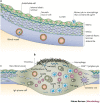
Figure 2. Proposal of events that lead to coronary-artery aneurysms in acute Kawasaki disease.
a | A small subset of circulating monocytes and/or macrophages contain the Kawasaki disease (KD) agent; these adhere to endothelial cells, and can enter the arterial wall at the intimal surface and through small arteries (vaso vasorum) in the adventitia. b | Infection of an artery leads to infiltration of additional circulating monocytes and/or macrophages. Macrophages secrete vascular endothelial growth factor (VEGF), matrix metalloproteinase 9 (MMP9), tumour necrosis factor-α (TNF-α) and other cytokines and enzymes. Antigens of the KD agent are processed by major histocompatibility complex class I. Antigen-specific CD8+ T lymphocytes target infected cells for destruction. Antigen-specific IgA B cells develop into plasma cells following exposure to local cytokines; specific antibody is produced to combat the agent. The intima is destroyed as endothelial cells become necrotic and are sloughed, and the thrombus adheres to this damaged surface. Subsequently, internal and external elastic laminae are fragmented, collagen fibres are disrupted and smooth muscle cells become necrotic; media and adventitia are no longer distinct. The structural integrity of the artery is then lost, and ballooning occurs. Myofibroblasts that secrete VEGF and MMP2 proliferate and can enter the organized thrombus, thereby forming neointima that can thicken over time. Neoangiogenesis in neointima and adventitia also occurs. c| The adjacent area of the artery that is not infiltrated by monocytes and/or macrophages that contain the KD agent is not affected.

References

    1. Suzuki A, et al. Coronary arterial lesions of Kawasaki disease: cardiac catheterization findings of 1100 cases. Pediatr. Cardiol. 1986;7:3–9. doi: 10.1007/BF02315475. - DOI - PubMed
    1. Newburger JW, et al. The treatment of Kawasaki syndrome with intravenous gammaglobulin. N. Engl. J. Med. 1986;315:341–347. doi: 10.1056/NEJM198608073150601. - DOI - PubMed
    1. Kato H, et al. Long-term consequences of Kawasaki disease. A 10- to 21-year follow-up study of 594 patients. Circulation. 1996;94:1379–1385. doi: 10.1161/01.CIR.94.6.1379. - DOI - PubMed
    1. Newburger JW, et al. Diagnosis, treatment, and long-term management of Kawasaki disease. Circulation. 2004;110:2747–2771. doi: 10.1161/01.CIR.0000145143.19711.78. - DOI - PubMed
    1. Landing BH, Larson EJ. Pathologic features of Kawasaki disease. Am. J. Cardiovasc. Pathol. 1987;1:218–229. - PubMed

Publication types

MeSH terms