. 2008 Mar 26;3(3):e1876.
doi: 10.1371/journal.pone.0001876.

Massive autophosphorylation of the Ser/Thr-rich domain controls protein kinase activity of TRPM6 and TRPM7

Affiliations

Massive autophosphorylation of the Ser/Thr-rich domain controls protein kinase activity of TRPM6 and TRPM7

Kristopher Clark et al. PLoS One. .

Abstract

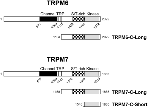
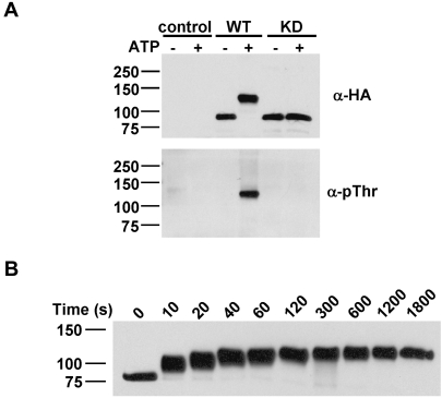
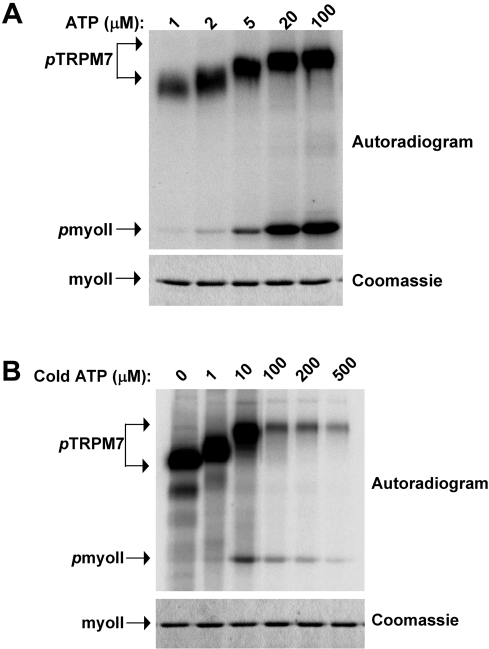
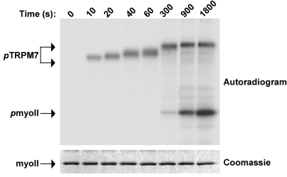
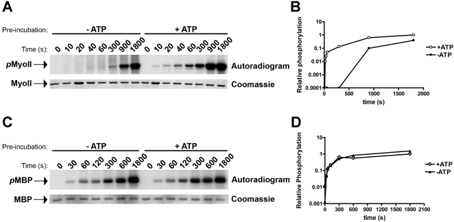
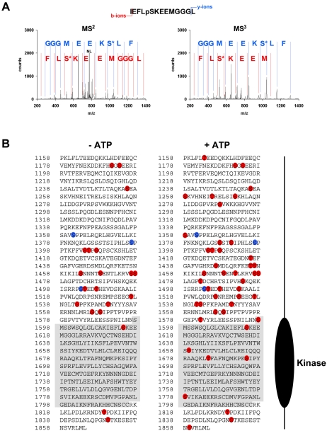
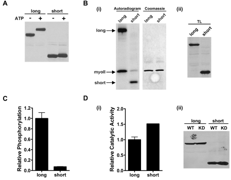
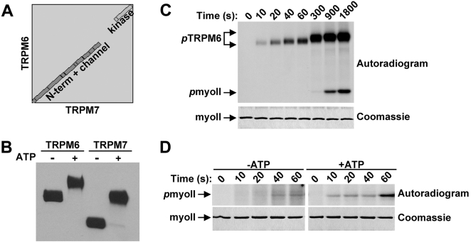
TRPM6 and TRPM7 are bifunctional proteins expressing a TRP channel fused to an atypical alpha-kinase domain. While the gating properties of TRPM6 and TRPM7 channels have been studied in detail, little is known about the mechanisms regulating kinase activity. Recently, we found that TRPM7 associates with its substrate myosin II via a kinase-dependent mechanism suggesting a role for autophosphorylation in substrate recognition. Here, we demonstrate that the cytosolic C-terminus of TRPM7 undergoes massive autophosphorylation (32+/-4 mol/mol), which strongly increases the rate of substrate phosphorylation. Phosphomapping by mass spectrometry indicates that the majority of autophosphorylation sites (37 out of 46) map to a Ser/Thr-rich region immediately N-terminal of the catalytic domain. Deletion of this region prevents substrate phosphorylation without affecting intrinsic catalytic activity suggesting that the Ser/Thr-rich domain contributes to substrate recognition. Surprisingly, the TRPM6-kinase is regulated by an analogous mechanism despite a lack of sequence conservation with the TRPM7 Ser/Thr-rich domain. In conclusion, our findings support a model where massive autophosphorylation outside the catalytic domain of TRPM6 and TRPM7 may facilitate kinase-substrate interactions leading to enhanced phosphorylation of those substrates.


Conflict of interest statement

Competing Interests: The authors have declared that no competing interests exist.

Figures

Figure 1
Figure 1. Schematic diagram depicting the constructs of TRPM6 and TRPM7 used in the current investigation.
TRPM6 and TRPM7 encode both a channel (black) and an α-kinase domain (grey). The typical TRP domain (stripes) is present directly C-terminal to the last transmembrane domain and a Ser/Thr-rich region (checkered) is present immediately N-terminal of the kinase domain. In addition to the full-length protein, the entire cytosolic C tail but excluding the TRP domain of TRPM6 and TRPM7 (TRPM6-C-long and TRPM7-C-long) as well as the isolated kinase domain of TRPM7 (TRPM7-C-short) were expressed as HA-tagged recombinant proteins in mammalian cells. Mouse and human TRPM7 sequences found in the NCBI database differ by a single amino acid. Since we used human TRPM7 in this study, we have corrected all positions to correspond to the human sequence and therefore Ser1511 and Ser1567 become Ser1512 and Ser1568, respectively.
Figure 2
Figure 2. TRPM7 undergoes extensive autophosphorylation of the C-terminus via a sequential series of phosphorylation events.
(A) Autophosphorylation of TRPM7 dramatically reduces its electrophoretic mobility. WT and KD TRPM7-C-long were incubated in the absence or presence of MnATP for 30 min and detected using anti-HA (top panel) and anti-pThr antibodies (bottom panel). (B) Kinetics of TRPM7 autophosphorylation. TRPM7-C-long was incubated with MnATP for the indicated times. Proteins were detected by immunoblotting using anti-HA antibodies.
Figure 3
Figure 3. Onset of substrate phosphorylation occurs after completion of TRPM7 autophosphorylation.
(A) Effect of reducing ATP concentration on TRPM7 autophosphorylation and myosin II phosphorylation. TRPM7-C-long was incubated with GST-myosin IIA in the presence of varying MnATP concentrations for 30 min at 30°C. The specific activity of ATP was maintained constant for all samples. The proteins were separated by SDS-PAGE, stained with coomassie brilliant blue (bottom panel) and phosphorylated proteins were detected by autoradiography (top panel). (B) Myosin II phosphorylation correlates with TRPM7 autophosphorylation even at exceedingly high specific activities. The experiment was performed as described in A except that the specific activity of the ATP solution was varied to detect myosin II phosphorylation at low ATP concentrations. All samples contained 5 µCi of [γ−32P]-ATP supplemented with the indicated concentrations of cold ATP.
Figure 4
Figure 4. TRPM7 autophosphorylation precedes phosphorylation of substrates.
TRPM7-C-long was incubated with GST-myosin IIB in the presence of Mn[γ−32P]-ATP for the indicated times. The proteins were separated by SDS-PAGE, revealed by coommassie staining (bottom panel) and phosphorylated proteins were detected by autoradiography (top panel).
Figure 5
Figure 5. Autophosphorylation of TRPM7 accelerates susbtrate phosphorylation without affecting catalytic activity.
(A) Kinetics of myosin II phosphorylation by TRPM7 prior to and after autophosphorylation. TRPM7-C-long was incubated in the absence (−ATP) or presence (+ATP) of cold MnATP to generate non-phosphorylated and completely autophosphorylated TRPM7. ATP was subsequently removed by washing the kinase with in vitro kinase buffer lacking ATP. Kinase reactions were initiated by adding Mn[γ−32P]-ATP and GST-myosin IIB and allowed to proceed at 30°C for the indicated times. Proteins were resolved by SDS-PAGE and visualized by coomassie staining (bottom panel). Phosphorylated myosin II (top panel) was detected by autoradiography. Note that the kinase pre-incubated in buffer without ATP (−ATP) must first complete autophosphorylation (5 min), apparent from the reduced electrophoretic mobility shift of TRPM7 (refer to Fig. 4), to acquire the ability to phosphorylate its substrates. (B) Quantification of myosin II phosphorylation by TRPM7 pre-incubated in absence (−ATP) or presence (+ATP) of ATP. 32P incorporation was measured by phosphorimaging analysis. The phosphorylation levels measured for myosin incubated for 30 min with TRPM7 that was pre-incubated with ATP was set to 1 and all other values are reported relative to that sample. (C) TRPM7 autophosphorylation does not affect catalytic activity. Experiment was performed as described in part A but using MBP as substrate. (D) Quantification of MBP phosphorylation by TRPM7 pre-incubated in absence (−ATP) or presence (+ATP) of ATP. Data was derived as described in part B.
Figure 6
Figure 6. Identification of phosphorylated residues in TRPM7 by LC-MS/MS.
WT and KD TRPM7-C-long were incubated in the presence of MnATP for 0 or 30 min. Tryptic peptides were analyzed using LTQ-FT mass spectrometer. (A) Representative MS2 and MS3 spectra for a TRPM7 peptide phosphorylated on Ser-1614. (m/z observed of parent ion was 823.3848, mass accuracy 1.85 ppm, +2 charge state; NL indicates neutral loss of H3PO4 which triggers acquisition of MS3 spectrum). The b+-ion series is indicated in red whereas the y+-ion series is in blue. S* refers to dehydroserine. (B) Diagram depicting the phosphorylated residues identified in WT (red) and KD (blue) TRPM7-C-long incubated in presence (right) or absence (left) of ATP.
Figure 7
Figure 7. Deletion of the Ser/Thr-rich domain in TRPM7 reduces substrate phosphorylation.
(A) Autophosphorylation of TRPM7-C-short only leads to a minor change in electrophoretic mobility on SDS-PAGE gels. TRPM7-C-short and TRPM7-C-long were incubated in the absence or presence of MnATP for 30 min at 30°C and subjected to immunoblotting using anti-HA antibodies. (B) TRPM7-C-short phosphorylates myosin II less efficiently than TRPM7-C-long. (i) TRPM7-C-short and TRPM7-C-long were incubated with GST-myosin IIB in the presence of Mn[γ−32P]-ATP. The proteins were separated by SDS-PAGE, revealed by coommassie staining (middle panel) and phosphorylated proteins were detected by autoradiography (left panel). (ii) Equal amounts of each kinase was verified by Western blotting (right panel). (C) Quantification of myosin II phosphorylation by TRPM7-C-short and TRPM7-C-long. 32P incorporation was measured by phosphorimaging analysis. The phosphorylation levels measured for myosin II incubated with TRPM7-C-long was set to 1 and values obtained for TRPM7-C-short are reported relative to that value. (D) Deletion of residues 1158-1548 does not affect catalytic activity. (i) TRPM7-C-long and TRPM7-C-short WT and KD were incubated with MH1 peptide in presence of Mn[γ−32P]-ATP. The level of phosphorylation was measured by scintillation counting. Background activity as determined using the KD mutant was substracted from the WT TRPM7 samples and catalytic activity was reported relative to that of TRPM7-C-long, which was set to 1. (ii) Equal amounts of each kinase were verified by Western blotting.
Figure 8
Figure 8. Regulation of TRPM6 by autophosphorylation.
(A) TRPM6 and TRPM7 display no sequence homology in the region spanning between the TRP box and the kinase domain. Alignment of amino acid sequences of TRPM6 and TRPM7 using Blast-2-Blast from NCBI. The gap is a result of low sequence similarity between the two proteins in the indicated region. (B) Autophosphorylation of TRPM6 leads to a dramatic shift in electrophoretic mobility on SDS-PAGE gels. TRPM6-C-long and TRPM7-C-long were incubated in the absence or presence of MnATP, resolved by SDS-PAGE and subsequently detected using anti-HA antibodies. (C) TRPM6 autophosphorylation precedes myosin II phosphorylation. TRPM6-C-long was incubated with GST-myosin IIB in the presence of Mn[γ−32P]-ATP for the indicated times. The proteins were separated by SDS-PAGE, revealed by coommassie staining (bottom panel) and phosphorylated proteins were detected by autoradiography (top panel). (D) Accelerated kinetics of myosin II phosphorylation after pre-incubation of TRPM6 with ATP. TRPM6-C-long was incubated in the absence or presence of MnATP to generate non-phosphorylated and completely autophosphorylated TRPM6. To initiate the phosphorylation of myosin II, in vitro kinase buffer containing Mn[γ−32P]-ATP and GST-myosin IIB was added. The reaction was allowed to proceed at 30°C and stopped by adding Laemmli buffer after the indicated times. Proteins were resolved by SDS-PAGE, visualized by coomassie staining (bottom panel) and phosphorylated proteins were detected by autoradiography (top panel).

References

    1. Bodding M. TRPM6: A Janus-like protein. Handb Exp Pharmacol. 2007;179:299–311. - PubMed
    1. Penner R, Fleig A. The Mg2+ and Mg(2+)-nucleotide-regulated channel-kinase TRPM7. Handb Exp Pharmacol. 2007;179:313–328. - PMC - PubMed
    1. Nadler MJ, Hermosura MC, Inabe K, Perraud AL, Zhu Q, et al. LTRPC7 is a Mg.ATP-regulated divalent cation channel required for cell viability. Nature. 2001;411:590–595. - PubMed
    1. Monteilh-Zoller MK, Hermosura MC, Nadler MJ, Scharenberg AM, Penner R, et al. TRPM7 provides an ion channel mechanism for cellular entry of trace metal ions. J Gen Physiol. 2003;121:49–60. - PMC - PubMed
    1. Voets T, Nilius B, Hoefs S, van der Kemp AW, Droogmans G, et al. TRPM6 forms the Mg2+ influx channel involved in intestinal and renal Mg2+ absorption. J Biol Chem. 2004;279:19–25. - PubMed

Publication types