Review
. 2008 May 1;100(1):1-18.
doi: 10.1002/bit.21838.

Medical biofilms

Affiliations
Review

Medical biofilms

James D Bryers. Biotechnol Bioeng. .

Abstract

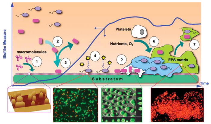
For more than two decades, Biotechnology and Bioengineering has documented research focused on natural and engineered microbial biofilms within aquatic and subterranean ecosystems, wastewater and waste-gas treatment systems, marine vessels and structures, and industrial bioprocesses. Compared to suspended culture systems, intentionally engineered biofilms are heterogeneous reaction systems that can increase reactor productivity, system stability, and provide inherent cell:product separation. Unwanted biofilms can create enormous increases in fluid frictional resistances, unacceptable reductions in heat transfer efficiency, product contamination, enhanced material deterioration, and accelerated corrosion. Missing from B&B has been an equivalent research dialogue regarding the basic molecular microbiology, immunology, and biotechnological aspects of medical biofilms. Presented here are the current problems related to medical biofilms; current concepts of biofilm formation, persistence, and interactions with the host immune system; and emerging technologies for controlling medical biofilms.


Figures

Figure 1
Figure 1
Three-dimensional reconstruction of a binary culture biofilm collected by confocal laser scanning microscopy. Biofilm z-direction “height” is 25 μm. The biofilm comprises the bacterial species, Klebsiella pneumoniae (green) and Pseudomonas aeruginosa (red), which have been visualized using species-specific monoclonal antibody-conjugated quantum dots (QDs). Unlike traditional fluorochrome stains, quantum dot luminescence is photostable and size tunable, allowing for multi-color emitted light from QDs of varying size all with a single excitation wavelength. Here, K. pneumoniae is labeled with monoclonal antibody-conjugated to CdSe-core, ZnS-shell nanocrystals (Green 560 nm emission maxima) and P. aeruginosa (red) monoclonal antibody-conjugated to CdSe/ZnS nanocrystals (Red 600 nm emission maxima).
Figure 2
Figure 2
Processes governing biofilm formation. Blue plot shows time course of net accumulation of biofilm on an initially clean substratum. Initially, ➀ substratum is biased either by naturally occurring macromolecular adsorption to surface or intentionally pre-treatment with molecules meant to attract mammalian cells. Suspended bacteria are then ➁ deposited at the substratum by a combination of many transport mechanisms. Cells may leave the surface or be permanently attached➂ soon after which they secrete quorum signals ➃ that initiate up-regulation of various genes (many related to virulence) on a community-wide basis. Attached cells secrete ➄ copious polymers (polysaccharides, proteins, oligonucleotides) forming a 3-D extracellular biofilm matrix. Biofilm continues to accumulate; ➅ consuming ambient nutrients, electron donors and acceptors, and attracting other bacterial species or mammalian cells. As a result of increases in shear stress or the onset of other cell: cell signaling events, portions or entire sections of biofilm can ➆ detach or slough off, to move downstream (if in the cardiovasculature, this is a deadly event known as a thromboembolism). Insets from left to right: atomic force microscope of protein pre-treatment of a glass substratum (Jarvis and Bryers, 2005); Live (green) Dead (red) Baclight stained adherent P. aeruginosa (Wagner and Bryers, 2004); CLSM image of a developing P. aeruginosa biofilm (TUDelft University); OCT gel-embedded P. aeruginosa biofilm stained with DAPI and CTC (courtesy G. McFeters and ASM.org image library).
Figure 3
Figure 3
Phenotypes of cells comprising the immune system.
Figure 4
Figure 4
Triggering dendritic cell response through TOL-like receptors and other adaptor proteins results in distinctive dendritic cell response depending on signal and activated receptor (from Pulendran et al., 2001).
Figure 5
Figure 5
Real-time monitoring of Staphylococcus aureus Xen29 in an experimental-rat endocarditis model. Two representative animals infected intravenously with either normal saline (control) or 106 CFU of S. aureus strain are shown. The animals were imaged ventrally, with their chest area shaved, to avoid background signal from animal hair. The process of infection was monitored daily by detecting photon emission around the region of interest (heart area) over a 6-day course (Xiong et al., 2005).
Figure 6
Figure 6
Hypothetical biomaterial engineered to enhance short and long-term infection immune response.
Figure 7
Figure 7
A: Effect of Ga on P. aeruginosa suspended growth. Ga(NO3)3 inhibits P. aeruginosa growth in a concentration-dependent manner. Experiments were performed in biofilm medium at 37°C, and data are the mean of four experiments; error bars indicate SEM. B: Ga prevents P. aeruginosa biofilm formation. Confocal microscopic images of GFP-expressing P. aeruginosa in flow cells perfused with biofilm medium without (left) and with (right) Ga, 5 days after inoculation. Experiments were performed at 25°C using 0.5 μg/mL Ga(NO3)3. Higher concentrations [1, 5, and 10 μM Ga(NO3)3] also inhibited biofilm formation. Top images are top-down views (x-y plane); bottom images are side views (x-z plane); the dotted line represents the biofilm growth surface; scale bars: 50 μm. Results are representative of six experiments (Kaneko et al., 2007).
Figure 8
Figure 8
A: Use of bi-specific fusion proteins to opsonize pathogenic bacteria and enhance phagocytosis. Human polymorphonuclear neutrophils (PMN) (B) and P. gingivalis (C) were incubated with different concentrations of BiFPs. This was followed by FITC-conjugated antimouse (filled circles) or FITC-conjugated antirabbit IgG (open circles) (Kobayashi et al., 2004).
Figure 9
Figure 9
Antigen presentation and pathways of vaccine response. Plasmid DNA or mRNA is taken up by dendritic cells for intracellular expression of antigen. Antigen can be secreted (not shown) and subsequently taken up by another DCs as an exogenous antigen. Antigen expressed intracellularly by a dendritic cell or taken up through cross-priming is presented by MHC-I to CD8+ T-cells (cytotoxic leukocytes; CTLs). Antigen taken in exogenously or directed by DNA or mRNA trafficking signals are processed by the MHC-II pathway and presented to CD4+ TH cells, which can subsequently secrete: soluble cytokine signals (e.g., IL-12) back to the dendritic cell, proliferative signals (e.g., IL-2 and IFN-γ) to Tc cells, or signals directed toward B-cells (e.g., IL-4) to induce B-cell proliferation and antibody secretion.

Similar articles

Cited by

References

    1. An YH, Friedman RJ. Concise review of mechanisms of bacterial adhesion to biomaterial surfaces. J Biomed Mater Res. 1998;43(3):338–348. - PubMed
    1. Anderson JM. Biological responses to materials. Annu Rev Mater Res. 2001;31:81–110.
    1. Anderson CF, Mosser DM. A novel phenotype for an activated macrophage: The type 2 activated macrophage. J Leukoc Biol. 2002;72:101–106. - PubMed
    1. Ankenbauer R, Sriyosachati S, Cox CD. Effects of siderophores on the growth of Pseudomonas aeruginosa in human serum and transferrin. Infect Immun. 1985;49:132–140. - PMC - PubMed
    1. Atkinson RA, Salah El Din AL, Kieffer B, Lefevre JF, Abdallah MA. Bacterial iron transport: 1H NMR determination of the three-dimensional structure of the gallium complex of pyoverdin G4R, the peptidic siderophore of Pseudomonas putida G4R. Biochemistry. 1998;37:15965–15973. - PubMed

MeSH terms