. 2008 Apr 22;47(16):4666-73.
doi: 10.1021/bi702130s. Epub 2008 Mar 27.

The first mutant of the Aequorea victoria green fluorescent protein that forms a red chromophore

Affiliations

The first mutant of the Aequorea victoria green fluorescent protein that forms a red chromophore

Alexander S Mishin et al. Biochemistry. .

Abstract

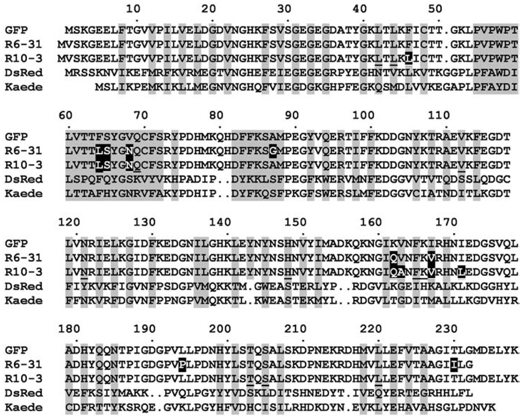
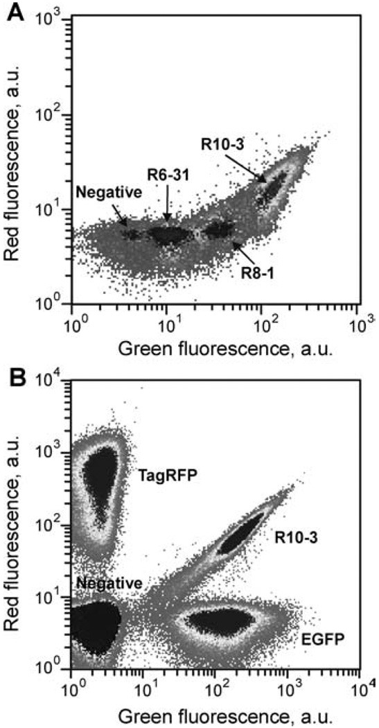
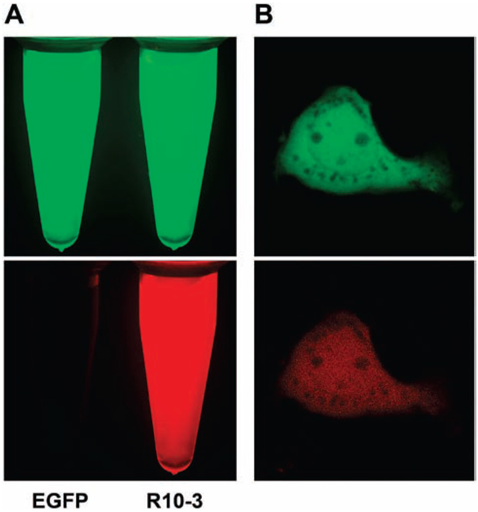
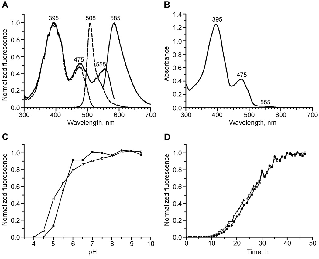
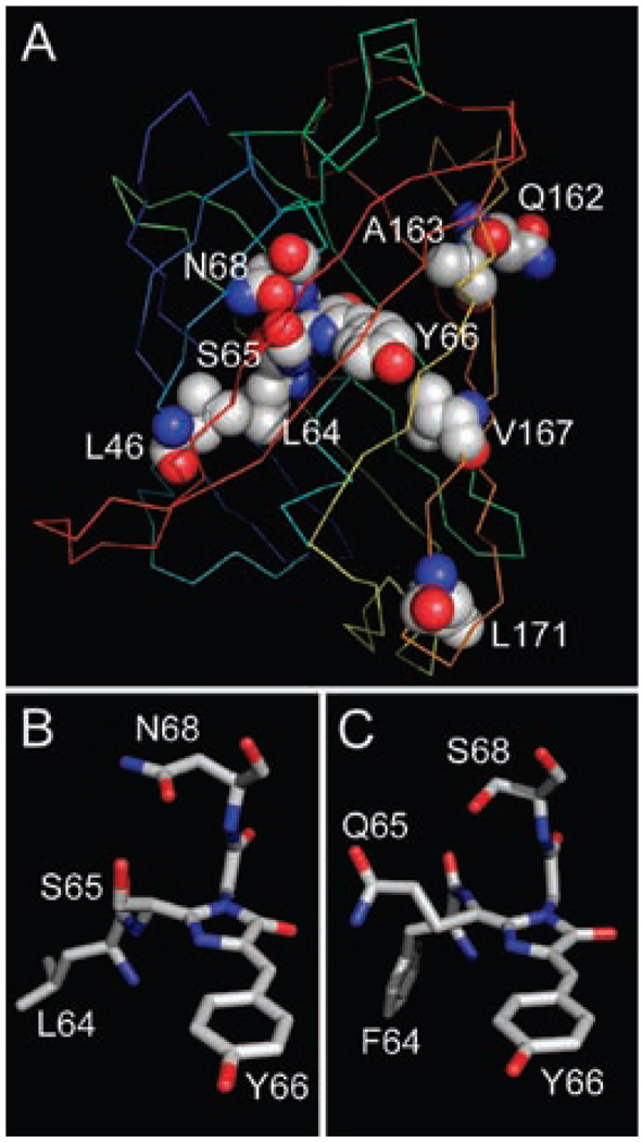
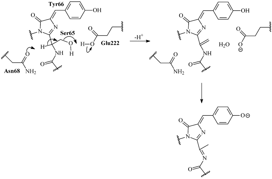
Green fluorescent protein (GFP) from a jellyfish, Aequorea victoria, and its mutants are widely used in biomedical studies as fluorescent markers. In spite of the enormous efforts of academia and industry toward generating its red fluorescent mutants, no GFP variants with emission maximum at more than 529 nm have been developed during the 15 years since its cloning. Here, we used a new strategy of molecular evolution aimed at generating a red-emitting mutant of GFP. As a result, we have succeeded in producing the first GFP mutant that substantially matures to the red-emitting state with excitation and emission maxima at 555 and 585 nm, respectively. A novel, nonoxidative mechanism for formation of the red chromophore in this mutant that includes a dehydration of the Ser65 side chain has been proposed. Model experiments showed that the novel dual-color GFP mutant with green and red emission is suitable for multicolor flow cytometry as an additional color since it is clearly separable from both green and red fluorescent tags.


Figures

FIGURE 1
FIGURE 1
Amino acid alignment of GFP, its mutants R6-31 and R10-3, Anthozoa red fluorescent protein DsRed, and green-to-red photoconvertible fluorescent protein Kaede. The residues whose side chains are in the interior of the β-can are shaded in gray. Introduced gaps are represented by dots. Substitutions in R6-31 and R10-3 are shaded in black. In R10-3, positions at which saturated mutagenesis was performed are underlined.
FIGURE 2
FIGURE 2
Flow cytometry analysis. The 488 nm line of argon and 568 nm line of krypton lasers were used to excite green and red fluorescence, respectively. (A) Comparison of bacterial cells with empty plasmid or expressing R6-31, R8-1, R10-3 mutants obtained consequently during molecular evolution. (B) Sorting of a mixture of bacterial cells expressing TagRFP, EGFP, R10-3, or empty plasmid. Note the clear separation of R10-3 from both EGFP and TagRFP.
FIGURE 3
FIGURE 3
Fluorescence microscopy of R10-3. (A) Comparison of EGFP (left tube) and R10-3 (right tube) fluorescence. Purified protein samples were photographed under a fluorescent stereomicroscope. Top: Excitation at 450–490 nm; detection above 510 nm (green channel). Bottom: Excitation at 540–580 nm; detection above 610 nm (red channel). (B) Confocal image of HEK293 cells expressing R10-3. Top: Excitation at 488 nm; detection at 500–530 nm (green channel). Bottom: Excitation at 543 nm; detection at 560–650 nm (red channel).
FIGURE 4
FIGURE 4
Spectral properties of the R10-3 mutant. (A) Excitation and emission spectra for green (dashed lines) and red (solid lines) fluorescence. Within each pair of lines the emission spectrum is the one at longer wavelengths. (B) Absorbance spectrum. (C) pH dependence of green (open squares) and red (filled circles) fluorescence. (D) Development of green (open squares) and red (filled circles) fluorescence in growing E. coli streak expressing R10-3.
FIGURE 5
FIGURE 5
Outline of the proposed three-dimensional structure of R10-3. (A) Ribbon scheme of the overall R10-3 structure modeled on the base of the GFP-F64 crystal structure (Protein Data Bank accession code 1EMM). The chromophore and residues, which are substituted in the R10-3 mutant, are labeled and shown in spacefilling representation. The protein backbone is colored from blue (N-end) to red (C-end). (B, C) Structural comparison of residues 64–68 in R10-3 (B) and DsRed (a monomer with presumably the catalytic conformation of Ser68 from Protein Data Bank accession code 1G7K) (C) shown in stick representation. Note the similar positioning of R10-3 Asn68 and DsRed Ser68 where oxygen atoms of the side chains are close to Cα at position 65. Amino acid substitutions followed by geometry optimization were performed using HyperChem software. Graphics are outputs of PyMOL software.
Scheme 1
Scheme 1
Proposed Nonoxidative Mechanism of the Red Chromophore Formation in R10-3a aDuring the first step, Asn68 and Glu222 promote dehydration of Ser65 resulting in a double bond between the Cα and Cβ atoms of Ser65. Then, an isomerization of this double bond and deprotonation of Tyr66 may lead to the DsRed-like chromophore where the GFP chromophore core is extended with the acylimine group.

Similar articles

Cited by

References

    1. Lippincott-Schwartz J, Patterson GH. Development and use of fluorescent protein markers in living cells. Science. 2003;300:87–91. - PubMed
    1. Verkhusha VV, Lukyanov KA. The molecular properties and applications of Anthozoa fluorescent proteins and chromoproteins. Nat. Biotechnol. 2004;22:289–296. - PubMed
    1. Chudakov DM, Lukyanov S, Lukyanov KA. Fluorescent proteins as a toolkit for in vivo imaging. Trends Biotechnol. 2005;23:605–613. - PubMed
    1. Johnson FH, Shimomura O, Saiga Y, Gershman LC, Reynolds GT, Waters JR. Quantum efficiency of Cypridina luminescence, with a note on that of Aequorea. J. Cell. Comp. Physiol. 1962;60:85–104.
    1. Chalfie M. Green fluorescent protein. Photochem. Photo-biol. 1995;62:651–656. - PubMed

Publication types

Substances